Application
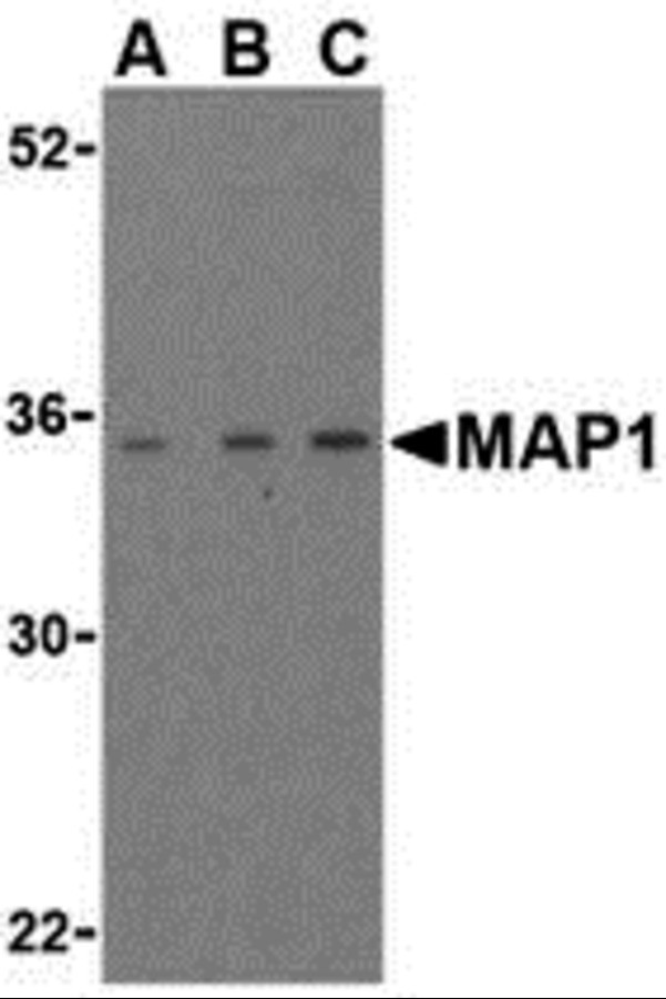
Western blot analysis of MAP-1 in EL4 cell lysate with MAP-1 antibody (IN) at (A) 1, (B) 2, and (C) 4 μg/mL.| Product Name | MAP1 Antibody |
|---|---|
| Antibody Type | Primary Antibodies |
| Product description | MAP1 Antibody: Apoptosis plays a major role in normal organism development, tissue homeostasis, and removal of damaged cells. Disruption of this process has been implicated in a variety of diseases such as cancer. Members of the Bcl-2 family are known to be critical regulators of this process. These proteins are characterized by the presence of several conserved motifs termed Bcl-2 homology (BH) domains. A related protein termed MAP-1 has recently been identified. This protein contains a BH3-like domain and induces caspase-dependent apoptosis in mammalian cells when overexpressed. It forms homodimers and associates with Bcl-2 family members such as Bax, Bcl-2, and Bcl-XL in vitro and in vivo. It has been suggested that MAP-1 associates with the tumor suppressor RASSF1A following death receptor activation, allowing a conformational change in Bax that leads to cellular apoptosis.1) Lockshin RA, Osborne B, and Zakeri Z. Cell death in the third millennium. Cell Death Differ. 2000; 7:2-7. |
| Immunogen | MAP-1 antibody was raised against a 15 amino acid synthetic peptide from near the center of human MAP-1. The immunogen is located within the last 50 amino acids of MAP1. |
| Clonality | Polyclonal |
|---|---|
| Isotype | IgG |
| Host Species | Rabbit |
| Tested Applications | ELISAWB |
| MAP1 antibody can be used for the detection of MAP-1 by Western blot at 1 to 4 μg/mL. | |
| Species Reactivity | HumanMouse |
| Concentration | 1mg/ml |
| Purification | Affinity purified |
| Gene Symbol | MOAP1 |
|---|---|
| Alternative Names | MAP1 Antibody: MAP-1 PNMA4 Modulator of apoptosis 1 Paraneoplastic antigen Ma4 MAP-1 |
Application
Western blot analysis of MAP-1 in EL4 cell lysate with MAP-1 antibody (IN) at (A) 1, (B) 2, and (C) 4 μg/mL.| Positive Control | 1) Cat. No. 1287 - EL4 Cell Lysate |
|---|---|
| Application Notes | MAP1 antibody can be used for the detection of MAP-1 by Western blot at 1 to 4 μg/mL. |
| Form | Liquid |
|---|---|
| Storage Instructions | MAP1 antibody can be stored at 4˚C for three months and -20˚C, stable for up to one year. As with all antibodies care should be taken to avoid repeated freeze thaw cycles. Antibodies should not be exposed to prolonged high temperatures. |
| Storage Buffer | MAP1 Antibody is supplied in PBS containing 0.02% sodium azide. |


