IHC
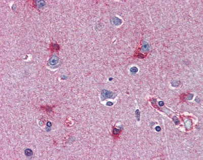
Human brain cortex tissue stained with MAT2A Antibody at 10 μg/mL followed by biotinylated anti-mouse IgG secondary antibody, alkaline phosphatase-streptavidin and chromogen.IHC
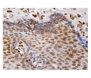
Immunoperoxidase staining of human esophageal tissue fixed in formalin and embedded in paraffin reveals nuclear staining of squamous epithelial cells.WB
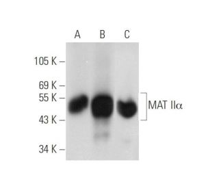
A: Control 293T Lysate was not transfected, B: Mouse MAT II alpha was transfected into 293T whole cell lysate, C: HeLa nuclear extract, Western blot analysis of MAT II alpha expression.| Product Name | MAT2A Antibody [AT3A2] |
|---|---|
| Antibody Type | Primary Antibodies |
| Immunogen | Recombinant human MAT2A (1-395aa) purified from E. coli |
| Clonality | Monoclonal |
|---|---|
| Isotype | IgG1, k |
| Host Species | Mouse |
| Tested Applications | IHCWB |
| MAT2A antibody can be used in Western Blot starting at 5 - 2 μg/mL, and immunohistochemistry starting at 20 μg/mL. | |
| Species Reactivity | HumanMouseRat |
| Concentration | 1mg/ml |
| Purification | Protein G |
| Gene Symbol | MAT2A |
|---|---|
| Alternative Names | MAT2A AdoMet synthase 2 AMS2 AdoMet synthetase 2 MATA2 MATII MAT 2 MAT-II SAMS2 |
IHC
Human brain cortex tissue stained with MAT2A Antibody at 10 μg/mL followed by biotinylated anti-mouse IgG secondary antibody, alkaline phosphatase-streptavidin and chromogen.
IHC
Immunoperoxidase staining of human esophageal tissue fixed in formalin and embedded in paraffin reveals nuclear staining of squamous epithelial cells.
WB
A: Control 293T Lysate was not transfected, B: Mouse MAT II alpha was transfected into 293T whole cell lysate, C: HeLa nuclear extract, Western blot analysis of MAT II alpha expression.| Application Notes | MAT2A antibody can be used in Western Blot starting at 5 - 2 μg/mL, and immunohistochemistry starting at 20 μg/mL. |
|---|
| Form | Liquid |
|---|---|
| Storage Instructions | MAT2A antibody can be stored short term 4 ˚C. For long term storage aliquot and store at -20 ˚C. As with all antibodies avoid freeze/thaw cycles. |
| Storage Buffer | PBS, pH 7.4, 0.1% sodium azide |


