Application
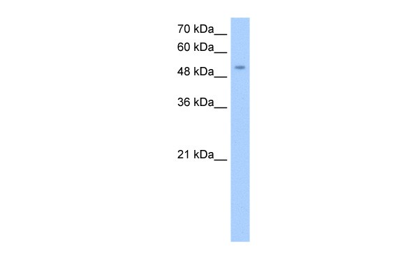
Antibody used in WB on Human HepG2 at 0.2-1 ug/ml.| Product Name | MFNG Antibody |
|---|---|
| Antibody Type | Primary Antibodies |
| Product description | MFNG is one of the evolutionarily conserved secreted proteins that act in the Notch receptor pathway to demarcate boundaries during embryonic development. Western blots using two different antibodies against two unique regions of this protein target confirm the same apparent molecular weight in our tests.This gene is a member of the fringe gene family which also includes Radical and Lunatic fringe. They all encode evolutionarily conserved secreted proteins that act in the Notch receptor pathway to demarcate boundaries during embryonic development. While their genomic structure is distinct from other glycosyltransferases, fringe proteins have a fucose-specific beta1,3 N-acetylglucosaminyltransferase activity that leads to elongation of O-linked fucose residues on Notch, which alters Notch signaling. Publication Note: This RefSeq record includes a subset of the publications that are available for this gene. Please see the Entrez Gene record to access additional publications.1) Veeraraghavalu, K., (2004) J. Virol. 78 (16), 8687-8700. |
| Immunogen | Antibody produced in rabbits immunized with a synthetic peptide corresponding a region of human MFNG. |
| Clonality | Polyclonal |
|---|---|
| Host Species | Rabbit |
| Tested Applications | WB |
| MFNG antibody can be used for detection of MFNG by ELISA at 1:312500. MFNG antibody can be used for detection of MFNG by western blot at 0.25 μg/mL, and HRP conjugated secondary antibody should be diluted 1:50,000 - 100,000. | |
| Species Reactivity | MouseRat |
| Concentration | 1mg/ml |
| Purification | Affinity purified |
| Gene Symbol | MFNG |
|---|---|
| Alternative Names | MFNG |
| Molecular Weight(MW) | 36 kDa |
Application
Antibody used in WB on Human HepG2 at 0.2-1 ug/ml.| Positive Control | 1) Cat. No. 1211 - HepG2 Cell Lysate |
|---|---|
| Application Notes | MFNG antibody can be used for detection of MFNG by ELISA at 1:312500. MFNG antibody can be used for detection of MFNG by western blot at 0.25 μg/mL, and HRP conjugated secondary antibody should be diluted 1:50,000 - 100,000. |
| Form | Liquid |
|---|---|
| Storage Instructions | For short periods of storage (days) store at 4˚C. For longer periods of storage, store MFNG antibody at -20˚C. As with any antibody avoid repeat freeze-thaw cycles. |
| Storage Buffer | Antibody is lyophilized in PBS buffer with 2% sucrose. Add 50 μL of distilled water. Final antibody concentration is 1 mg/mL. |


