_1.jpg)
_1.jpg)
Application
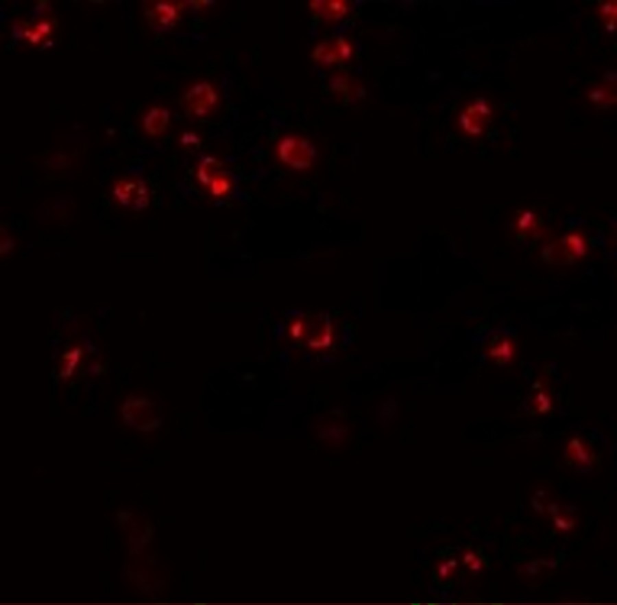
Immunofluorescence of NALP3 in K562 cells with NALP3 antibody at 20 μg/mL.Application
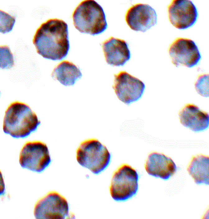
Immunocytochemistry of NALP3 in K562 cells with NALP3 antibody at 2 μg/mL.Application
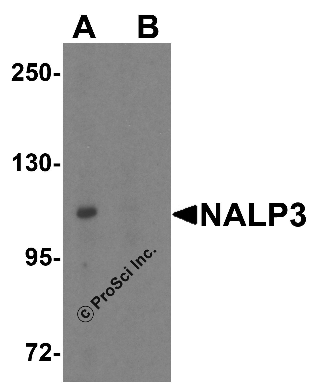
Western blot analysis of NALP3 in K562 cell lysate with NALP3 antibody at 1 μg/mL (A) in the absence and (B) in the presence of blocking peptide.IHC
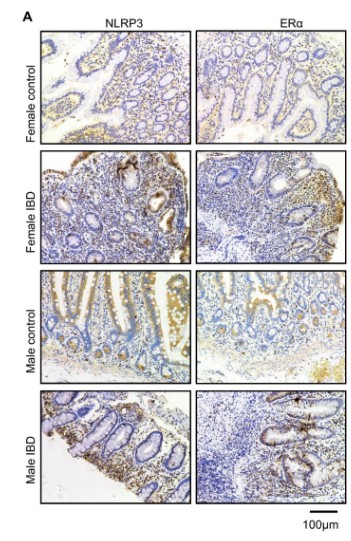
(A) Immunohistochemistry analysis of NLRP3 and ERα in different intestinal tissue sections. (Scale bars, 100µm).WB
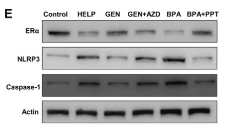
(E) Protein expression levels of ERα, NLRP3, and Caspase-1 of LMH cells. (F–H) Quantified protein level of ERα, NLRP3, and Caspase-1 (normalized to β-actin). *P < 0.05, **P < 0.01, ***P < 0.001.WB
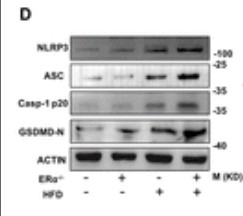
(D) Protein expressions of NLRP3 inflammasome components and GSDMD-N of WT and ERα–/– mice.WB
(B) Protein expressions of NLRP3 inflammasome components and GSDMD-N.WB
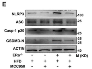
(E) Protein expressions of NLRP3 inflammasome components and GSDMD-N.WB
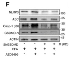
(F) Immunoblot analysis of NLRP3 inflammasome components and GSDMD-N.WB
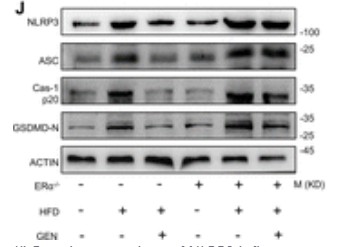
(J) Protein expressions of NLRP3 inflammasome components and GSDMD-N.WB
(C) Protein levels of NLRP3, ASC and Caspase-1 in colon tissues.WB
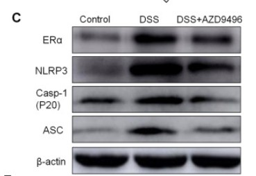
(C) Protein levels of NLRP3, ASC, Caspase-1 and ERα in colon tissues.WB
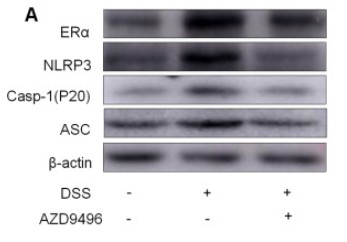
(A) Protein levels of NLRP3, ASC and Caspase-1 in colon tissues.IF
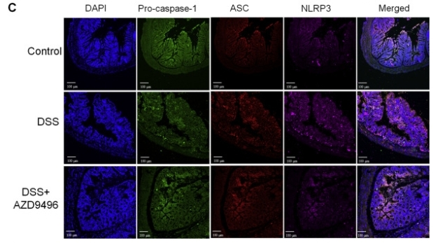
(C) Colon tissue was immunofluorescently stained with Cy5-NLRP3 antibody, PE-ASC antibody and FITC-pro-Caspase-1 antibody.| Product Name | NALP3 Antibody |
|---|---|
| Antibody Type | Primary Antibodies |
| Product description | NALP3 Antibody: NALP3, a member of Nod-like receptors, has a crucial role in inflammation and immunity and may be a proximal sensor of cellular stress and danger signals. NALP3 forms a caspase-1 activating molecular complex termed the inflammasome. The inflammasome allows activation of IL-1β, the key player of the inflammation and fever. NALP3 gene encodes a pyrin like protein which contains a pyrin domain, a nucleotide binding site (NBS) domain, and a leucine rich repeat (LRR) motif. NALP3 protein interacts with apoptosis associated speck like protein containing a CARD and may function as an inducer of apoptosis and an activator of NF-κB signaling. Defects in NALP3 have been associated with familial cold autoinflammatory syndrome (FCAS), Muckle-Wells syndrome (MWS), chronic infantile neurological cutaneous and articular (CINCA) syndrome, and neonatal-onset multisystem inflammatory disease (NOMID).1) Chamaillard M, Girardin SE, Viala J, et al. Nods, Nalps and Naip: intracellular regulators of bacterial-induced inflammation. Cell. Microbiol.2003; 5:581-92. |
| Immunogen | NALP3 antibody was raised against a 16 amino acid synthetic peptide from near the amino terminus of human NALP3. The immunogen is located within amino acids 120 - 170 of NALP3. |
| Clonality | Polyclonal |
|---|---|
| Isotype | IgG |
| Host Species | Rabbit |
| Tested Applications | ELISAICCIFIHCWB |
| NALP3 antibody can be used for detection of NALP3 by Western blot at 1 μg/mL. Antibody can also be used for Immunocytochemistry starting at 2 μg/mL. For immunofluorescence start at 20 μg/mL. | |
| Species Reactivity | HumanMouse |
| Concentration | 1mg/ml |
| Purification | Affinity purified |
| Gene Symbol | NLRP3 |
|---|---|
| Alternative Names | NALP3 Antibody: AII AVP FCU MWS FCAS CIAS1 NALP3 C1orf7 CLR1.1 PYPAF1 AGTAVPRL NACHT LRR and PYD domains-containing protein 3 Cold autoinflammatory syndrome 1 protein |
| Molecular Weight(MW) | Predicted: 101, 114 kDa Observed: 110 kDa |
Application
Immunofluorescence of NALP3 in K562 cells with NALP3 antibody at 20 μg/mL.
Application
Immunocytochemistry of NALP3 in K562 cells with NALP3 antibody at 2 μg/mL.
Application
Western blot analysis of NALP3 in K562 cell lysate with NALP3 antibody at 1 μg/mL (A) in the absence and (B) in the presence of blocking peptide.
IHC
(A) Immunohistochemistry analysis of NLRP3 and ERα in different intestinal tissue sections. (Scale bars, 100µm).
WB
(E) Protein expression levels of ERα, NLRP3, and Caspase-1 of LMH cells. (F–H) Quantified protein level of ERα, NLRP3, and Caspase-1 (normalized to β-actin). *P < 0.05, **P < 0.01, ***P < 0.001.
WB
(D) Protein expressions of NLRP3 inflammasome components and GSDMD-N of WT and ERα–/– mice.
WB
(B) Protein expressions of NLRP3 inflammasome components and GSDMD-N.
WB
(E) Protein expressions of NLRP3 inflammasome components and GSDMD-N.
WB
(F) Immunoblot analysis of NLRP3 inflammasome components and GSDMD-N.
WB
(J) Protein expressions of NLRP3 inflammasome components and GSDMD-N.
WB
(C) Protein levels of NLRP3, ASC and Caspase-1 in colon tissues.
WB
(C) Protein levels of NLRP3, ASC, Caspase-1 and ERα in colon tissues.
WB
(A) Protein levels of NLRP3, ASC and Caspase-1 in colon tissues.
IF
(C) Colon tissue was immunofluorescently stained with Cy5-NLRP3 antibody, PE-ASC antibody and FITC-pro-Caspase-1 antibody.| Positive Control | K562 Cell Lysate |
|---|---|
| Application Notes | NALP3 antibody can be used for detection of NALP3 by Western blot at 1 μg/mL. Antibody can also be used for Immunocytochemistry starting at 2 μg/mL. For immunofluorescence start at 20 μg/mL. |
| Form | Liquid |
|---|---|
| Storage Instructions | NALP3 antibody can be stored at 4˚C for three months and -20˚C, stable for up to one year. As with all antibodies care should be taken to avoid repeated freeze thaw cycles. Antibodies should not be exposed to prolonged high temperatures. |
| Storage Buffer | NALP3 Antibody is supplied in PBS containing 0.02% sodium azide. |


