Application
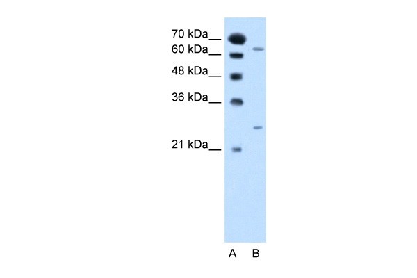
Antibody used in WB on Human Jurkat 2.5 ug/ml.Application
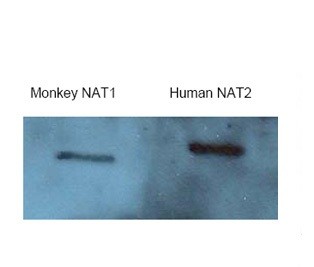
Antibody used in WB.WB
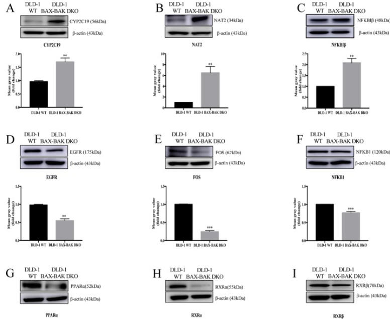
(A–L) Cropped images showed protein expression levels of CYP2C19, NAT2, NFKBIβ, EGFR, FOS, NFKB1, PPARα, RXRα, RXRβ, TPMT, UGCG, and XPC in DLD-1 WT and DLD-1 BAX–BAK DKO cells. Bar charts presented relative changes of mean gray value, which was normalized to that of β-actin.WB
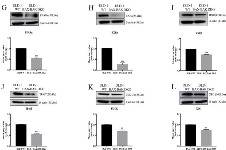
(A–L) Cropped images showed protein expression levels of CYP2C19, NAT2, NFKBIβ, EGFR, FOS, NFKB1, PPARα, RXRα, RXRβ, TPMT, UGCG, and XPC in DLD-1 WT and DLD-1 BAX–BAK DKO cells. Bar charts presented relative changes of mean gray value, which was normalized to that of β-actin.| Product Name | NAT2 Antibody |
|---|---|
| Antibody Type | Primary Antibodies |
| Product description | NAT2 is a N-acetyltransferase 2 (arylamine N-acetyltransferase 2). This enzyme functions to both activate and deactivate arylamine and hydrazine drugs and carcinogens. Polymorphisms in its gene are reponsible for the N-acetylation polymorphism in which human populations segregate into rapid,intermediate, and slow acetylator phenotypes. Polymorphisms in NAT2 are also associated with higher incidences of cancer and drug toxicity.This gene encodes N-acetyltransferase 2 (arylamine N-acetyltransferase 2). This enzyme functions to both activate and deactivate arylamine and hydrazine drugs and carcinogens. Polymorphisms in this gene are reponsible for the N-acetylation polymorphism in which human populations segregate into rapid,intermediate, and slow acetylator phenotypes. Polymorphisms in NAT2 are also associated with higher incidences of cancer and drug toxicity. A second arylamine N-acetyltransferase gene (NAT1) is located near NAT2.1) Tamer, L., (2006) Cell Biochem. Funct. 24 (2), 131-135. |
| Immunogen | Antibody produced in rabbits immunized with a synthetic peptide corresponding a region of human NAT2. |
| Clonality | Polyclonal |
|---|---|
| Host Species | Rabbit |
| Tested Applications | ELISAWB |
| NAT2 antibody can be used for detection of NAT2 by ELISA at 1:312500. NAT2 antibody can be used for detection of NAT2 by western blot at 2.5 μg/mL, and HRP conjugated secondary antibody should be diluted 1:50,000 - 100,000. | |
| Species Reactivity | Human |
| Concentration | 1mg/ml |
| Purification | Protein A |
| Gene Symbol | NAT2 |
|---|---|
| Alternative Names | NAT2 AAC2 PNAT NAT-2 |
| Molecular Weight(MW) | 32 kDa |
Application
Antibody used in WB on Human Jurkat 2.5 ug/ml.
Application
Antibody used in WB.
WB
(A–L) Cropped images showed protein expression levels of CYP2C19, NAT2, NFKBIβ, EGFR, FOS, NFKB1, PPARα, RXRα, RXRβ, TPMT, UGCG, and XPC in DLD-1 WT and DLD-1 BAX–BAK DKO cells. Bar charts presented relative changes of mean gray value, which was normalized to that of β-actin.
WB
(A–L) Cropped images showed protein expression levels of CYP2C19, NAT2, NFKBIβ, EGFR, FOS, NFKB1, PPARα, RXRα, RXRβ, TPMT, UGCG, and XPC in DLD-1 WT and DLD-1 BAX–BAK DKO cells. Bar charts presented relative changes of mean gray value, which was normalized to that of β-actin.| Positive Control | 1) Cat. No. 1205 - Jurkat Cell Lysate |
|---|---|
| Application Notes | NAT2 antibody can be used for detection of NAT2 by ELISA at 1:312500. NAT2 antibody can be used for detection of NAT2 by western blot at 2.5 μg/mL, and HRP conjugated secondary antibody should be diluted 1:50,000 - 100,000. |
| Form | Liquid |
|---|---|
| Storage Instructions | For short periods of storage (days) store at 4˚C. For longer periods of storage, store NAT2 antibody at -20˚C. As with any antibody avoid repeat freeze-thaw cycles. |
| Storage Buffer | IgG in phosphate buffered saline , pH 7.4, 150mM NaCl, 0.02% sodium azide and 50% glycerol.Store at -20 °C.Stable for 12 months from date of receipt |


