Application
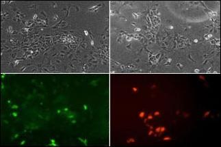
ES cells were transiently transfected with flag-tagged mouse NeuroD1 (tagged on N-term).Fixed 24h post transfection.Stained for flag tag (red) to check that some cells express protein. Most protein was in nucleus but some was cytoplasmic. Stained with NeuroD1 N-term antibodies at 1:100. NeuroD1 N-term antibody showed strong and clear staining with similar pattern to the flag staining.(Supplied byApplication
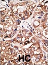
Formalin-fixed and paraffin-embedded human cancer tissue reacted with the primary antibody, which was peroxidase-conjugated to the secondary antibody, followed by DAB staining. BC = breast carcinoma; HC = hepatocarcinoma.Application
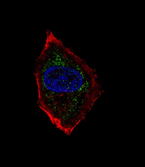
Fluorescent confocal image of HepG2 cell stained with hNeuroD1-Q30. HepG2 cells were fixed with 4% PFA (20 min), permeabilized with Triton X-100 (0.1%, 10 min), then incubated with hNeuroD1-Q30 primary antibody (1:25). For secondary antibody, Alexa Fluor 488 conjugated donkey anti-rabbit antibody (green) was used (1:400).Cytoplasmic actin was counterstained with Alexa Fluor 555 (red) conjugated PhApplication
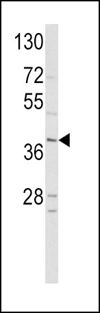
Western blot analysis of hNeuroD1-Q30 in HepG2 cell line lysates (35ug/lane)| Product Name | NeuroD1 Antibody |
|---|---|
| Antibody Type | Primary Antibodies |
| Antigen Alias | Neurogenic differentiation factor 1, NeuroD, NeuroD1, Class A basic helix-loop-helix protein 3, bHLHa3, NEUROD1, BHLHA3, NEUROD |
| Product description | NeuroD1 acts as a differentiation factor during neurogenesis. They are expressed transiently in a subset of neurons in the central and peripheral nervous systems at the time of their terminal differentiation. NeuroD1 is a basic helix-loop-helix (bHLH) protein contain 1 bHLH domain. NeuroD1 is a transcriptional activator, for efficient DNA binding it requires dimerization with another bHLH protein. It was reported that NeuroD1 involves heterodimerization with the ubiquitous bHLH protein E47, and regulates insulin gene expression by binding to a critical E-box motif on the insulin promoter. Defects in NEUROD1 causes maturity onset diabetes of the young type VI. MODY6 is a form of non-insulin-dependent diabetes mellitus (NIDDM) characterized by an autosomal dominant mode of inheritance, onset during young adulthood and a primary defect in insulin secretion.1) Breslin, M.B., et al., J. Biol. Chem. 278(40):38991-38997 (2003). |
| Immunogen | This NeuroD1 antibody is generated from rabbits immunized with a KLH conjugated synthetic peptide between 15-45 amino acids from the N-terminal region of human NeuroD1. |
| Clonality | Polyclonal |
|---|---|
| Isotype | Ig |
| Host Species | Rabbit |
| Tested Applications | IFIHC-PWB |
| For WB starting dilution is: 1:1000 For IF starting dilution is: 1:10~50 For IHC-P starting dilution is: 1:50~100: |
|
| Species Reactivity | Human |
| Concentration | 1mg/ml |
| Gene Symbol | NEUROD1 |
|---|---|
| Alternative Names | Neurogenic differentiation factor 1 NeuroD NeuroD1 Class A basic helix-loop-helix protein 3 bHLHa3 NEUROD1 BHLHA3 NEUROD |
| Molecular Weight(MW) | 40 kDa |
Application
ES cells were transiently transfected with flag-tagged mouse NeuroD1 (tagged on N-term).Fixed 24h post transfection.Stained for flag tag (red) to check that some cells express protein. Most protein was in nucleus but some was cytoplasmic. Stained with NeuroD1 N-term antibodies at 1:100. NeuroD1 N-term antibody showed strong and clear staining with similar pattern to the flag staining.(Supplied by
Application
Formalin-fixed and paraffin-embedded human cancer tissue reacted with the primary antibody, which was peroxidase-conjugated to the secondary antibody, followed by DAB staining. BC = breast carcinoma; HC = hepatocarcinoma.
Application
Fluorescent confocal image of HepG2 cell stained with hNeuroD1-Q30. HepG2 cells were fixed with 4% PFA (20 min), permeabilized with Triton X-100 (0.1%, 10 min), then incubated with hNeuroD1-Q30 primary antibody (1:25). For secondary antibody, Alexa Fluor 488 conjugated donkey anti-rabbit antibody (green) was used (1:400).Cytoplasmic actin was counterstained with Alexa Fluor 555 (red) conjugated Ph
Application
Western blot analysis of hNeuroD1-Q30 in HepG2 cell line lysates (35ug/lane)| Application Notes | For WB starting dilution is: 1:1000 For IF starting dilution is: 1:10~50 For IHC-P starting dilution is: 1:50~100: |
|---|
| Form | Liquid |
|---|---|
| Storage Instructions | Store at 4˚C for three months and -20˚C, stable for up to one year. As with all antibodies care should be taken to avoid repeated freeze thaw cycles. Antibodies should not be exposed to prolonged high temperatures. |
| Storage Buffer | Supplied in PBS with 0.09% (W/V) sodium azide. |


