Application
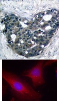
Top Image: Immunohistochemical analysis of paraffin-embedded human breast carcinoma tissue using p56Dok-2 (Ab-299). Bottom Image: Immunofluorescence staining of methanol-fixed HeLa cells using p56Dok-2 (Ab-299).Application
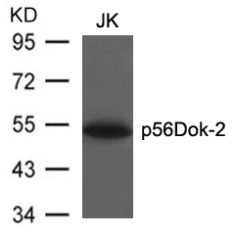
Western blot analysis of lysed extracts from JK cells using p56Dok-2 (Ab-299).| Product Name | p56Dok 2 Antibody |
|---|---|
| Antibody Type | Primary Antibodies |
| Antigen Alias | p56DOK, p56dok-2, CHK1, DOK2 |
| Product description | DOK proteins are enzymatically inert adaptor or scaffolding proteins. They provide a docking platform for the assembly of multimolecular signaling complexes. DOK2 may modulate the cellular proliferation induced by IL-4, as well as IL-2 and IL-3. May be involved in modulating Bcr-Abl signaling. Attenuates EGF-stimulated MAP kinase activation1) Feng Cong, et, al. (1999) Mol. Cell. Biol; 19: 8314 - 8325. Serge Lemay, et, al. (2000) Mol. Cell. Biol; 20: 2743 - 2754. Ute Schaeper, et, al. (2000) J. Cell Biol; 149: 1419. Miyuki Honma, et, al. (2006) Genes Cells; 11: 143 - 151. |
| Immunogen | p56Dok-2 (Ab-299) antibody was raised against a peptide sequence around aa.297~301 (G-E-Y-A-V) derived from Human p56Dok-2. |
| Clonality | Polyclonal |
|---|---|
| Host Species | Rabbit |
| Tested Applications | IFIHCWB |
| Western Blot: 1:500~1:1000, Immunohistochemistry: 1:50~1:100, Immunofluorescence: 1:100~1:200: |
|
| Species Reactivity | Human |
| Concentration | 1mg/ml |
| Purification | Affinity purified |
| Gene Symbol | DOK2 |
|---|---|
| Alternative Names | p56DOK p56dok-2 CHK1 DOK2 |
| Molecular Weight(MW) | 56 kDa |
| Tissue Specificity | This antibody detects endogenous level of total p56Dok-2 protein. |
| Entrez Gene | 9046 |
|---|---|
| Protein Accession | NP_003965.2 |
Application
Top Image: Immunohistochemical analysis of paraffin-embedded human breast carcinoma tissue using p56Dok-2 (Ab-299). Bottom Image: Immunofluorescence staining of methanol-fixed HeLa cells using p56Dok-2 (Ab-299).
Application
Western blot analysis of lysed extracts from JK cells using p56Dok-2 (Ab-299).| Application Notes | Western Blot: 1:500~1:1000, Immunohistochemistry: 1:50~1:100, Immunofluorescence: 1:100~1:200: |
|---|
| Form | Liquid |
|---|---|
| Storage Instructions | Store antibody at -20˚C for up to one year. |
| Storage Buffer | Antibody supplied in phosphate buffered saline (without Mg2+ and Ca2+), pH 7.4, 150mM NaCl, 0.02% sodium azide and 50% glycerol. |


