Application
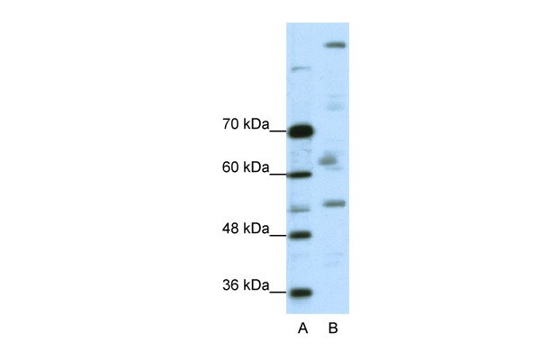
Antibody used in WB on Human A204 cells at 0.2-1 ug/ml.| Product Name | PEG3 Antibody |
|---|---|
| Antibody Type | Primary Antibodies |
| Antigen Alias | PEG3, PW1, ZNF904, ZSCAN24, ZKSCAN22 |
| Product description | PEG3 induces apoptosis in cooperation with SIAH1A and acts as a mediator between TP53/p53 and BAX in a neuronal death pathway that is activated by DNA damage. PEG3 acts synergistically with TRAF2 and inhibits TNF induced apoptosis through activation of NF-kappa-B. PEG3 also possesses a tumor suppressing activity in glioma cells.1) Dowdy, S.C., et al., (2005) Gynecol. Oncol. 99 (1), 126-134. |
| Immunogen | Antibody produced in rabbits immunized with a synthetic peptide corresponding a region of human PEG3. |
| Clonality | Polyclonal |
|---|---|
| Host Species | Rabbit |
| Tested Applications | ELISAWB |
| PEG3 antibody can be used for detection of PEG3 by ELISA at 1:312500. PEG3 antibody can be used for detection of PEG3 by western blot at 0.25 μg/mL, and HRP conjugated secondary antibody should be diluted 1:50,000 - 100,000.: |
|
| Species Reactivity | Human |
| Concentration | 1mg/ml |
| Purification | Affinity purified |
| Gene Symbol | PEG3 |
|---|---|
| Alternative Names | PEG3 PW1 ZNF904 ZSCAN24 ZKSCAN22 |
| Molecular Weight(MW) | 181 kDa |
Application
Antibody used in WB on Human A204 cells at 0.2-1 ug/ml.| Positive Control | 1) A204 Cell Lysate |
|---|---|
| Application Notes | PEG3 antibody can be used for detection of PEG3 by ELISA at 1:312500. PEG3 antibody can be used for detection of PEG3 by western blot at 0.25 μg/mL, and HRP conjugated secondary antibody should be diluted 1:50,000 - 100,000.: |
| Form | Liquid |
|---|---|
| Storage Instructions | For short periods of storage (days) store at 4˚C. For longer periods of storage, store PEG3 antibody at -20˚C. As with any antibody avoid repeat freeze-thaw cycles. |
| Storage Buffer | Antibody is lyophilized in PBS buffer with 2% sucrose. Add 50 μL of distilled water. Final antibody concentration is 1 mg/mL. |


