Application
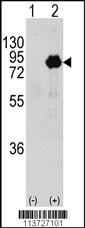
Western blot analysis of PIK3R2 using rabbit polyclonal PIK3R2 Antibody (T447) using 293 cell lysates (2 ug/lane) either nontransfected (Lane 1) or transiently transfected with the PIK3R2 gene (Lane 2).| Product Name | PIK3R2 Antibody |
|---|---|
| Antibody Type | Primary Antibodies |
| Antigen Alias | Phosphatidylinositol 3-kinase regulatory subunit beta, PI3-kinase regulatory subunit beta, PI3K regulatory subunit beta, PtdIns-3-kinase regulatory subunit beta, Phosphatidylinositol 3-kinase 85 kDa regulatory subunit beta, PI3-kinase subunit p85-beta, Ptd |
| Product description | Protein kinases are enzymes that transfer a phosphate group from a phosphate donor, generally the g phosphate of ATP, onto an acceptor amino acid in a substrate protein. By this basic mechanism, protein kinases mediate most of the signal transduction in eukaryotic cells, regulating cellular metabolism, transcription, cell cycle progression, cytoskeletal rearrangement and cell movement, apoptosis, and differentiation. The family has been classified into 8 major groups based on sequence comparison of their tyrosine (PTK) or serine/threonine (STK) kinase catalytic domains. PIK3R2 binds to activated Protein Tyrosine Kinases, which are phosphorylated, through its SH2 domain, and acts as an adaptor, mediating the association of the P110 catalytic unit to the plasma membrane.1) Khan, N.A., et al., J. Neurovirol. 9(6):584-593 (2003). |
| Immunogen | This PIK3R2 antibody is generated from rabbits immunized with a KLH conjugated synthetic peptide between 442-471 amino acids from human PIK3R2. |
| Clonality | Polyclonal |
|---|---|
| Isotype | Ig |
| Host Species | Rabbit |
| Tested Applications | WB |
| For WB starting dilution is: 1:1000: |
|
| Species Reactivity | Human |
| Concentration | 1mg/ml |
| Purification | Affinity purified |
| Gene Symbol | PIK3R2 |
|---|---|
| Alternative Names | Phosphatidylinositol 3-kinase regulatory subunit beta PI3-kinase regulatory subunit beta PI3K regulatory subunit beta PtdIns-3-kinase regulatory subunit beta Phosphatidylinositol 3-kinase 85 kDa regulatory subunit beta PI3-kinase subunit p85-beta Ptd |
| Molecular Weight(MW) | 82 kDa |
| Sequence Similarities | Predicted species reactivity based on immunogen sequence: Bovine |
Application
Western blot analysis of PIK3R2 using rabbit polyclonal PIK3R2 Antibody (T447) using 293 cell lysates (2 ug/lane) either nontransfected (Lane 1) or transiently transfected with the PIK3R2 gene (Lane 2).| Application Notes | For WB starting dilution is: 1:1000: |
|---|
| Form | Liquid |
|---|---|
| Storage Instructions | Store at 4˚C for three months and -20˚C, stable for up to one year. As with all antibodies care should be taken to avoid repeated freeze thaw cycles. Antibodies should not be exposed to prolonged high temperatures. |
| Storage Buffer | Supplied in PBS with 0.09% (W/V) sodium azide. |


