Application
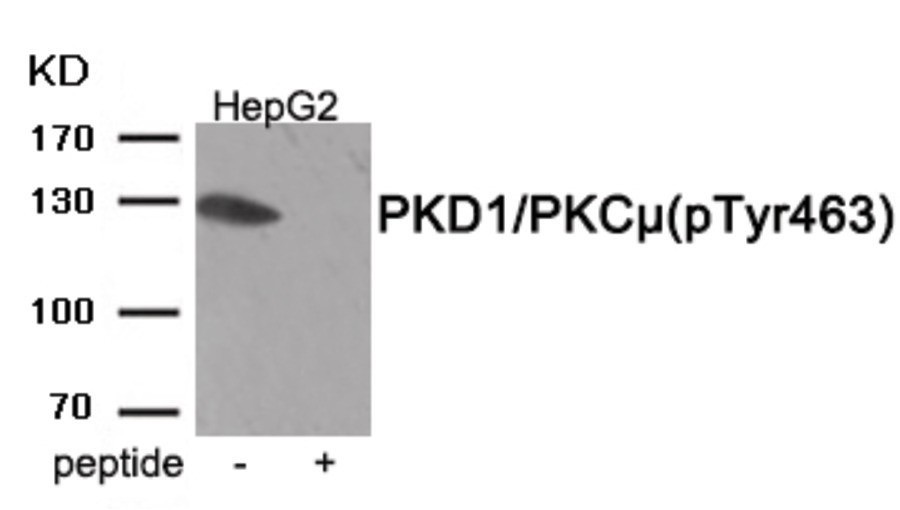
Western blot analysis of extracts from HepG2 tissue using PKD1/PKC μ (Phospho-Tyr463) Antibody. The lane on the right is treated with the antigen-specific peptide.| Product Name | PKD1/PKC μ (phospho Tyr463) Antibody |
|---|---|
| Antibody Type | Primary Antibodies |
| Antigen Alias | KPCD1, PKC-mu, PKCM, PKD, PRKD1 |
| Product description | Serine/threonine-protein kinase that converts transient diacylglycerol (DAG) signals into prolonged physiological effects downstream of PKC, and is involved in the regulation of MAPK8/JNK1 and Ras signaling, Golgi membrane integrity and trafficking, cell survival through NF-kappa-B activation, cell migration, cell differentiation by mediating HDAC7 nuclear export, cell proliferation via MAPK1/3 (ERK1/2) signaling, and plays a role in cardiac hypertrophy, VEGFA-induced angiogenesis, genotoxic-induced apoptosis and flagellin-stimulated inflammatory response. Phosphorylates the epidermal growth factor receptor (EGFR) on dual threonine residues, which leads to the suppression of epidermal growth factor (EGF)-induced MAPK8/JNK1 activation and subsequent JUN phosphorylation. Phosphorylates RIN1, inducing RIN1 binding to 14-3-3 proteins YWHAB, YWHAE and YWHAZ and increased competition with RAF1 for binding to GTP-bound form of Ras proteins (NRAS, HRAS and KRAS). Acts downstream of the heterotrimeric G-protein beta/gamma-subunit complex to maintain the structural integrity of the Golgi membranes, and is required for protein transport along the secretory pathway. In the trans-Golgi network (TGN), regulates the fission of transport vesicles that are on their way to the plasma membrane. May act by activating the lipid kinase phosphatidylinositol 4-kinase beta (PI4KB) at the TGN for the local synthesis of phosphorylated inositol lipids, which induces a sequential production of DAG, phosphatidic acid (PA) and lyso-PA (LPA) that are necessary for membrane fission and generation of specific transport carriers to the cell surface. Under oxidative stress, is phosphorylated at Tyr-463 via SRC-ABL1 and contributes to cell survival by activating IKK complex and subsequent nuclear translocation and activation of NFKB1. Involved in cell migration by regulating integrin alpha-5/beta-3 recycling and promoting its recruitment in newly forming focal adhesion.1) Döppler H, Storz P (2007)J Biol Chem 282, 31873-81. Qin L, Zeng H, Zhao D (2006)J Biol Chem 281, 32550-8. Storz P, Döppler H, Toker A (2004)Mol Pharmacol 66, 870-9 . |
| Immunogen | PKD1/PKC μ (phospho Tyr463) antibody was raised against a peptide sequence around phosphorylation site of Tyrosine463 (R-Y-Y (p)-K-E) derived from Human PKD1/PKC μ. |
| Modification | p-Tyr463 |
| Clonality | Polyclonal |
|---|---|
| Isotype | IgG |
| Host Species | Rabbit |
| Tested Applications | WB |
| Western Blot: 1:500~1:1000: |
|
| Species Reactivity | HumanMouseRat |
| Concentration | 1mg/ml |
| Purification | Affinity purified |
| Gene Symbol | PRKD1 |
|---|---|
| Alternative Names | KPCD1 PKC-mu PKCM PKD PRKD1 |
| Molecular Weight(MW) | 130 kDa |
| Tissue Specificity | The antibody detects endogenous level of PKD1/PKC μ only when phosphorylated at tyrosine 463. |
| Entrez Gene | 5587 |
|---|---|
| Protein Accession | NP_002733.2 |
Application
Western blot analysis of extracts from HepG2 tissue using PKD1/PKC μ (Phospho-Tyr463) Antibody. The lane on the right is treated with the antigen-specific peptide.| Application Notes | Western Blot: 1:500~1:1000: |
|---|
| Form | Liquid |
|---|---|
| Storage Instructions | Store antibody at -20˚C for up to one year. |
| Storage Buffer | Antibody supplied in phosphate buffered saline (without Mg2+ and Ca2+), pH 7.4, 150mM NaCl, 0.02% sodium azide and 50% glycerol. |


