Application
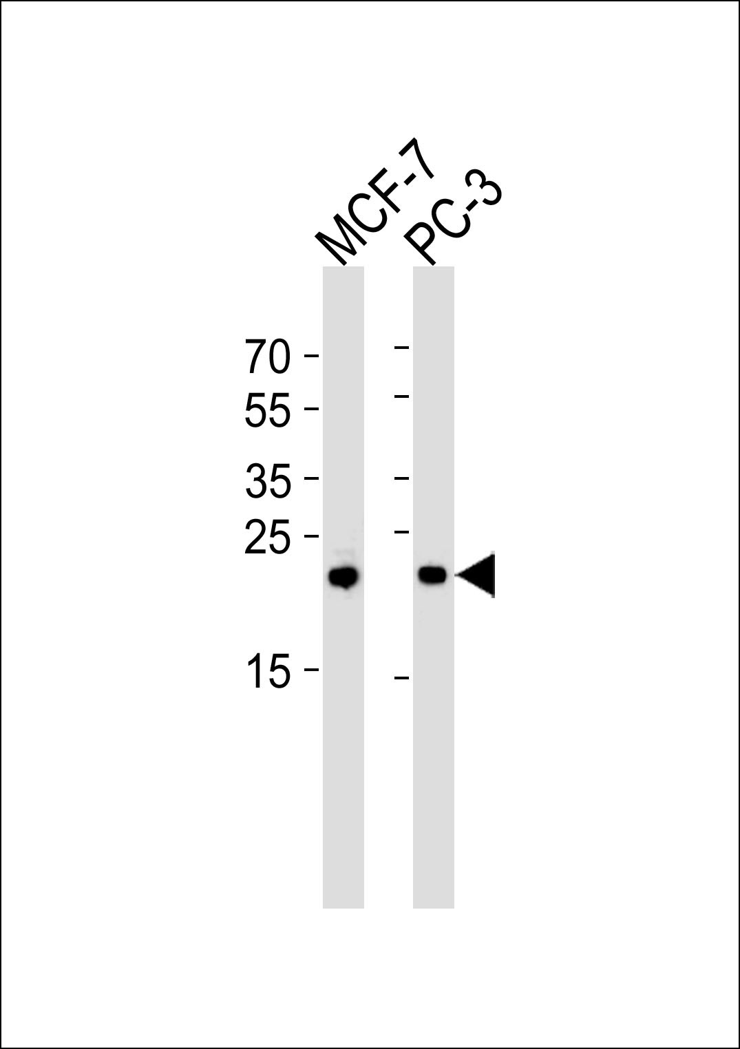
Western blot analysis of lysates from MCF-7, PC-3 cell line (from left to right), using PSMD10 Antibody at 1:1000 at each lane.Application
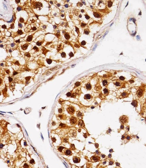
Immunohistochemical analysis of paraffin-embedded H.testis section using PSMD10 Antibody . Antibody was diluted at 1:25 dilution. A peroxidase-conjugated goat anti-rabbit IgG at 1:400 dilution was used as the secondary antibody, followed by DAB staining.Application
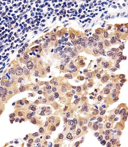
Immunohistochemical analysis of paraffin-embedded H.lung adenocarcinoma section using PSMD10 Antibody . Antibody was diluted at 1:25 dilution. A peroxidase-conjugated goat anti-rabbit IgG at 1:400 dilution was used as the secondary antibody, followed by DAB staining.Application
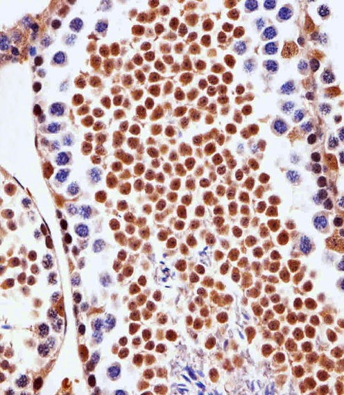
Immunohistochemical analysis of paraffin-embedded M.testis section using PSMD10 Antibody . Antibody was diluted at 1:25 dilution. A peroxidase-conjugated goat anti-rabbit IgG at 1:400 dilution was used as the secondary antibody, followed by DAB staining.Application
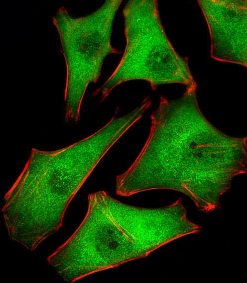
Fluorescent image of Hela cells stained with PSMD10 Antibody . Antibody was diluted at 1:25 dilution. An Alexa Fluor 488-conjugated goat anti-rabbit lgG at 1:400 dilution was used as the secondary antibody (green). Cytoplasmic actin was counterstained with Alexa Fluor 555 conjugated with Phalloidin (red).| Product Name | PSMD10 Antibody |
|---|---|
| Antibody Type | Primary Antibodies |
| Antigen Alias | 26S proteasome non-ATPase regulatory subunit 10, 26S proteasome regulatory subunit p28, Gankyrin, p28(GANK), PSMD10 |
| Product description | Acts as a chaperone during the assembly of the 26S proteasome, specifically of the PA700/19S regulatory complex (RC). In the initial step of the base subcomplex assembly is part of an intermediate PSMD10:PSMC4:PSMC5:PAAF1 module which probably assembles with a PSMD5:PSMC2:PSMC1:PSMD2 module. Independently of the proteasome, regulates EGF-induced AKT activation through inhibition of the RHOA/ROCK/PTEN pahway, leading to prolonged AKT activation. Plays an important role in RAS-induced tumorigenesis.1) Hori T.,et al.Gene 216:113-122(1998). |
| Immunogen | This PSMD10 antibody is generated from a rabbit immunized with a KLH conjugated synthetic peptide between 43-76 amino acids from the Central region of human PSMD10. |
| Clonality | Polyclonal |
|---|---|
| Isotype | Ig |
| Host Species | Rabbit |
| Tested Applications | IFIHC-PWB |
| For IF starting dilution is: 1:25 For IHC-P starting dilution is: 1:25 For WB starting dilution is: 1:1000: |
|
| Species Reactivity | Human |
| Concentration | 1mg/ml |
| Purification | Affinity purified |
| Gene Symbol | PSMD10 |
|---|---|
| Alternative Names | 26S proteasome non-ATPase regulatory subunit 10 26S proteasome regulatory subunit p28 Gankyrin p28(GANK) PSMD10 |
| Molecular Weight(MW) | 24 kDa |
| Sequence Similarities | Predicted species reactivity based on immunogen sequence: Mouse |
Application
Western blot analysis of lysates from MCF-7, PC-3 cell line (from left to right), using PSMD10 Antibody at 1:1000 at each lane.
Application
Immunohistochemical analysis of paraffin-embedded H.testis section using PSMD10 Antibody . Antibody was diluted at 1:25 dilution. A peroxidase-conjugated goat anti-rabbit IgG at 1:400 dilution was used as the secondary antibody, followed by DAB staining.
Application
Immunohistochemical analysis of paraffin-embedded H.lung adenocarcinoma section using PSMD10 Antibody . Antibody was diluted at 1:25 dilution. A peroxidase-conjugated goat anti-rabbit IgG at 1:400 dilution was used as the secondary antibody, followed by DAB staining.
Application
Immunohistochemical analysis of paraffin-embedded M.testis section using PSMD10 Antibody . Antibody was diluted at 1:25 dilution. A peroxidase-conjugated goat anti-rabbit IgG at 1:400 dilution was used as the secondary antibody, followed by DAB staining.
Application
Fluorescent image of Hela cells stained with PSMD10 Antibody . Antibody was diluted at 1:25 dilution. An Alexa Fluor 488-conjugated goat anti-rabbit lgG at 1:400 dilution was used as the secondary antibody (green). Cytoplasmic actin was counterstained with Alexa Fluor 555 conjugated with Phalloidin (red).| Application Notes | For IF starting dilution is: 1:25 For IHC-P starting dilution is: 1:25 For WB starting dilution is: 1:1000: |
|---|
| Form | Liquid |
|---|---|
| Storage Instructions | Store at 4˚C for three months and -20˚C, stable for up to one year. As with all antibodies care should be taken to avoid repeated freeze thaw cycles. Antibodies should not be exposed to prolonged high temperatures. |
| Storage Buffer | Supplied in PBS with 0.09% (W/V) sodium azide. |


