Application
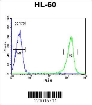
Flow cytometric analysis of HL-60 cells (right histogram) compared to a negative control cell (left histogram).FITC-conjugated goat-anti-rabbit secondary antibodies were used for the analysis.Application
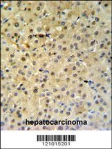
PSME1 Antibody IHC analysis in formalin fixed and paraffin embedded hepatocarcinoma followed by peroxidase conjugation of the secondary antibody and DAB staining.Application
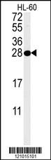
Western blot analysis in HL-60 cell line lysates (35ug/lane). This demonstrates the PSME1 antibody detected the PSME1 protein (arrow).| Product Name | PSME1 Antibody |
|---|---|
| Antibody Type | Primary Antibodies |
| Antigen Alias | Proteasome activator complex subunit 1, 11S regulator complex subunit alpha, REG-alpha, Activator of multicatalytic protease subunit 1, Interferon gamma up-regulated I-5111 protein, IGUP I-5111, Proteasome activator 28 subunit alpha, PA28a, PA28alpha, PSME |
| Product description | The 26S proteasome is a multicatalytic proteinase complex with a highly ordered structure composed of 2 complexes, a 20S core and a 19S regulator. The 20S core is composed of 4 rings of 28 non-identical subunits; 2 rings are composed of 7 alpha subunits and 2 rings are composed of 7 beta subunits. The 19S regulator is composed of a base, which contains 6 ATPase subunits and 2 non-ATPase subunits, and a lid, which contains up to 10 non-ATPase subunits. Proteasomes are distributed throughout eukaryotic cells at a high concentration and cleave peptides in an ATP/ubiquitin-dependent process in a non-lysosomal pathway. An essential function of a modified proteasome, the immunoproteasome, is the processing of class I MHC peptides. The immunoproteasome contains an alternate regulator, referred to as the 11S regulator or PA28, that replaces the 19S regulator. Three subunits (alpha, beta and gamma) of the 11S regulator have been identified. This gene encodes the alpha subunit of the 11S regulator, one of the two 11S subunits that is induced by gamma-interferon. Three alpha and three beta subunits combine to form a heterohexameric ring.1) Ruckrich, T., et al. Leukemia 23(6):1098-1105(2009) Lemaire, R., et al. J. Proteome Res. 6(11):4127-4134(2007) Kobayashi, K., et al. Brain Res. 1170, 129-139 (2007) |
| Immunogen | This PSME1 antibody is generated from rabbits immunized with a KLH conjugated synthetic peptide between 220-249 amino acids from the C-terminal region of human PSME1. |
| Clonality | Polyclonal |
|---|---|
| Isotype | Ig |
| Host Species | Rabbit |
| Tested Applications | FACSIHC-PWB |
| For WB starting dilution is: 1:1000 For IHC-P starting dilution is: 1:50~100 For FACS starting dilution is: 1:10~50: |
|
| Species Reactivity | Human |
| Concentration | 1mg/ml |
| Purification | Affinity purified |
| Gene Symbol | PSME1 |
|---|---|
| Alternative Names | Proteasome activator complex subunit 1 11S regulator complex subunit alpha REG-alpha Activator of multicatalytic protease subunit 1 Interferon gamma up-regulated I-5111 protein IGUP I-5111 Proteasome activator 28 subunit alpha PA28a PA28alpha PSME |
| Molecular Weight(MW) | 29 kDa |
| Sequence Similarities | Predicted species reactivity based on immunogen sequence: Bovine |
Application
Flow cytometric analysis of HL-60 cells (right histogram) compared to a negative control cell (left histogram).FITC-conjugated goat-anti-rabbit secondary antibodies were used for the analysis.
Application
PSME1 Antibody IHC analysis in formalin fixed and paraffin embedded hepatocarcinoma followed by peroxidase conjugation of the secondary antibody and DAB staining.
Application
Western blot analysis in HL-60 cell line lysates (35ug/lane). This demonstrates the PSME1 antibody detected the PSME1 protein (arrow).| Application Notes | For WB starting dilution is: 1:1000 For IHC-P starting dilution is: 1:50~100 For FACS starting dilution is: 1:10~50: |
|---|
| Form | Liquid |
|---|---|
| Storage Instructions | Store at 4˚C for three months and -20˚C, stable for up to one year. As with all antibodies care should be taken to avoid repeated freeze thaw cycles. Antibodies should not be exposed to prolonged high temperatures. |
| Storage Buffer | Supplied in PBS with 0.09% (W/V) sodium azide. |


