Application
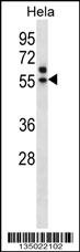
Western blot analysis in Hela cell line lysates (35ug/lane).Application
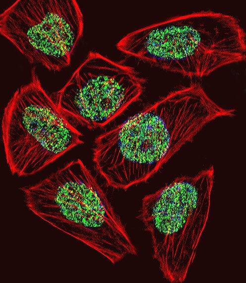
Fluorescent confocal image of U251 cell stained with TADA3L Antibody .U251 cells were fixed with 4% PFA (20 min), permeabilized with Triton X-100 (0.1%, 10 min), then incubated with TADA3L primary antibody (1:25). For secondary antibody, Alexa Fluor 488 conjugated donkey anti-rabbit antibody (green) was used (1:400).Cytoplasmic actin was counterstained with Alexa Fluor 555 (red) conjugated Phalloi| Product Name | TADA3L Antibody |
|---|---|
| Antibody Type | Primary Antibodies |
| Antigen Alias | Transcriptional adapter 3, ADA3 homolog, hADA3, STAF54, Transcriptional adapter 3-like, ADA3-like protein, TADA3, ADA3, TADA3L |
| Product description | Many DNA-binding transcriptional activator proteins enhance the initiation rate of RNA polymerase II-mediated gene transcription by interacting functionally with the general transcription machinery bound at the basal promoter. Adaptor proteins are usually required for this activation, possibly to acetylate and destabilize nucleosomes, thereby relieving chromatin constraints at the promoter. The protein encoded by this gene is a transcriptional activator adaptor and has been found to be part of the PCAF histone acetylase complex. In addition, it associates with the tumor suppressor protein p53 and is required for full activity of p53 and p53-mediated apoptosis. At least four alternatively spliced variants have been found for this gene, but the full-length nature of some variants has not been determined. [provided by RefSeq].1) Bailey, S.D., et al. Diabetes Care 33(10):2250-2253(2010) |
| Immunogen | This TADA3L antibody is generated from rabbits immunized with a KLH conjugated synthetic peptide between 386-414 amino acids from the C-terminal region of human TADA3L. |
| Clonality | Polyclonal |
|---|---|
| Isotype | Ig |
| Host Species | Rabbit |
| Tested Applications | IFWB |
| For WB starting dilution is: 1:1000 For IF starting dilution is: 1:10~50: |
|
| Species Reactivity | Human |
| Concentration | 1mg/ml |
| Purification | Affinity purified |
| Gene Symbol | TADA3 |
|---|---|
| Alternative Names | Transcriptional adapter 3 ADA3 homolog hADA3 STAF54 Transcriptional adapter 3-like ADA3-like protein TADA3 ADA3 TADA3L |
| Molecular Weight(MW) | 49 kDa |
| Sequence Similarities | Predicted species reactivity based on immunogen sequence: Xenopus |
Application
Western blot analysis in Hela cell line lysates (35ug/lane).
Application
Fluorescent confocal image of U251 cell stained with TADA3L Antibody .U251 cells were fixed with 4% PFA (20 min), permeabilized with Triton X-100 (0.1%, 10 min), then incubated with TADA3L primary antibody (1:25). For secondary antibody, Alexa Fluor 488 conjugated donkey anti-rabbit antibody (green) was used (1:400).Cytoplasmic actin was counterstained with Alexa Fluor 555 (red) conjugated Phalloi| Application Notes | For WB starting dilution is: 1:1000 For IF starting dilution is: 1:10~50: |
|---|
| Form | Liquid |
|---|---|
| Storage Instructions | Store at 4˚C for three months and -20˚C, stable for up to one year. As with all antibodies care should be taken to avoid repeated freeze thaw cycles. Antibodies should not be exposed to prolonged high temperatures. |
| Storage Buffer | Supplied in PBS with 0.09% (W/V) sodium azide. |


