WB
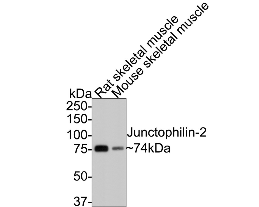
Western blot analysis of Junctophilin-2 on different lysates with Rabbit anti-Junctophilin-2 antibody at 1/2,000 dilution. Lane 1: Rat skeletal muscle tissue lysate, Lane 2: Mouse skeletal muscle tissue lysate, Lysates/proteins at 20 µg/Lane. Exposure time: 2 minutes; 8% SDS-PAGE gel. Proteins were transferred to a PVDF membrane and blocked with 5% NFDM/TBST for 1 hour at room temperature. The primary antibody at 1/2,000 dilution was used in 5% NFDM/TBST at room temperature for 2 hours. Goat Anti-Rabbit IgG - HRP Secondary Antibody at 1:300,000 dilution was used for 1 hour at room temperature.IHC
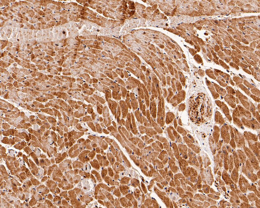
Immunohistochemical analysis of paraffin-embedded human heart tissue with Rabbit anti-Junctophilin-2 antibody at 1/100 dilution. The section was pre-treated using heat mediated antigen retrieval with Tris-EDTA buffer (pH 9.0) for 20 minutes. The tissues were blocked in 1% BSA for 20 minutes at room temperature, washed with ddH2O and PBS, and then probed with the primary antibody at 1/100 dilution for 1 hour at room temperature. The detection was performed using an HRP conjugated compact polymer system. DAB was used as the chromogen. Tissues were counterstained with hematoxylin and mounted with DPX.IHC
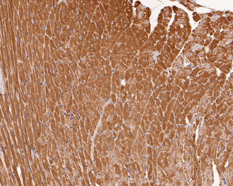
Immunohistochemical analysis of paraffin-embedded mouse heart tissue with Rabbit anti-Junctophilin-2 antibody at 1/400 dilution. The section was pre-treated using heat mediated antigen retrieval with Tris-EDTA buffer (pH 9.0) for 20 minutes. The tissues were blocked in 1% BSA for 20 minutes at room temperature, washed with ddH2O and PBS, and then probed with the primary antibody at 1/400 dilution for 1 hour at room temperature. The detection was performed using an HRP conjugated compact polymer system. DAB was used as the chromogen. Tissues were counterstained with hematoxylin and mounted with DPX.IHC
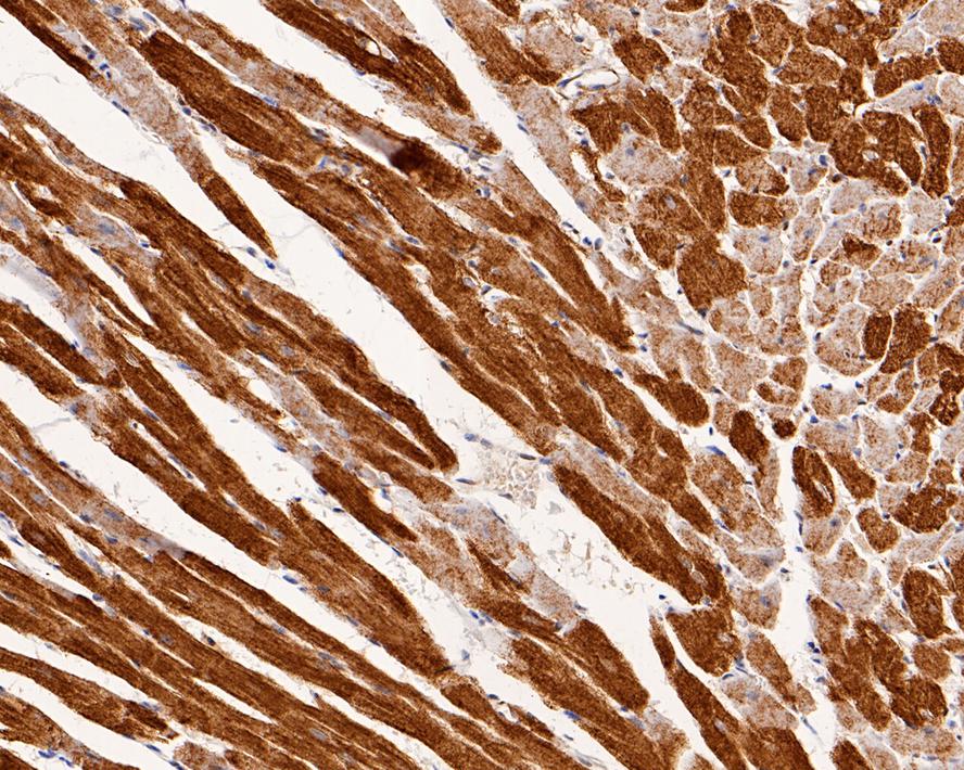
Immunohistochemical analysis of paraffin-embedded rat heart tissue with Rabbit anti-Junctophilin-2 antibody at 1/400 dilution. The section was pre-treated using heat mediated antigen retrieval with Tris-EDTA buffer (pH 9.0) for 20 minutes. The tissues were blocked in 1% BSA for 20 minutes at room temperature, washed with ddH2O and PBS, and then probed with the primary antibody at 1/400 dilution for 1 hour at room temperature. The detection was performed using an HRP conjugated compact polymer system. DAB was used as the chromogen. Tissues were counterstained with hematoxylin and mounted with DPX.FC
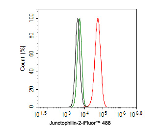
Flow cytometric analysis of C2C12 cells labeling Junctophilin-2. Cells were fixed and permeabilized. Then stained with the primary antibody (1ug/ml) (red) compared with Rabbit IgG Isotype Control (green). After incubation of the primary antibody at +4℃ for an hour, the cells were stained with a 488 conjugate-Goat anti-Rabbit IgG Secondary antibody at 1/1,000 dilution for 30 minutes at +4℃. Unlabelled sample was used as a control (cells without incubation with primary antibody; black).| Product Name | Junctophilin-2 Rabbit Polyclonal Antibody |
|---|---|
| Antibody Type | Primary Antibodies |
| Product description | Junctophilin-2 (JPH2) is a cardiac specific member of the junctophilins, a newly characterized family of junctional membrane complex proteins important in physically approximating the plasmalemmal L-type calcium channel and the sarcoplasmic reticulum ryanodine receptor for calcium-induced calcium release. It acts probably by anchoring the plasma membrane and endoplasmic reticulum. JPH2 contributes to the construction of skeletal muscle triad junctions, and plays an essential role in heart development. |
| Immunogen | Synthetic peptide within Human Junctophilin-2 aa 418-467 / 696. |
| Clonality | Polyclonal |
|---|---|
| Isotype | IgG |
| Host Species | Rabbit |
| Tested Applications | FCIHCWB |
| WB:1:2000 IHC:1:100-1:400 FC:1:50-1:1000 |
|
| Species Reactivity | HumanMouseRat |
| Concentration | 1mg/ml |
| Purification | Affinity purified |
| Alternative Names | FLJ40969 antibody JP-2 antibody JP2 antibody Jph2 antibody JPH2_HUMAN antibody Junctophilin 2 antibody Junctophilin type 2 antibody Junctophilin-2 antibody OTTHUMP00000031651 antibody OTTHUMP00000031652 antibody |
|---|---|
| Molecular Weight(MW) | 74 kDa |
| Cellular Localization | Cell membrane, Sarcoplasmic reticulum membrane, Endoplasmic reticulum membrane; Nucleus. |
| SwissProt ID | Q9BR39 |
|---|
WB
Western blot analysis of Junctophilin-2 on different lysates with Rabbit anti-Junctophilin-2 antibody at 1/2,000 dilution. Lane 1: Rat skeletal muscle tissue lysate, Lane 2: Mouse skeletal muscle tissue lysate, Lysates/proteins at 20 µg/Lane. Exposure time: 2 minutes; 8% SDS-PAGE gel. Proteins were transferred to a PVDF membrane and blocked with 5% NFDM/TBST for 1 hour at room temperature. The primary antibody at 1/2,000 dilution was used in 5% NFDM/TBST at room temperature for 2 hours. Goat Anti-Rabbit IgG - HRP Secondary Antibody at 1:300,000 dilution was used for 1 hour at room temperature.
IHC
Immunohistochemical analysis of paraffin-embedded human heart tissue with Rabbit anti-Junctophilin-2 antibody at 1/100 dilution. The section was pre-treated using heat mediated antigen retrieval with Tris-EDTA buffer (pH 9.0) for 20 minutes. The tissues were blocked in 1% BSA for 20 minutes at room temperature, washed with ddH2O and PBS, and then probed with the primary antibody at 1/100 dilution for 1 hour at room temperature. The detection was performed using an HRP conjugated compact polymer system. DAB was used as the chromogen. Tissues were counterstained with hematoxylin and mounted with DPX.
IHC
Immunohistochemical analysis of paraffin-embedded mouse heart tissue with Rabbit anti-Junctophilin-2 antibody at 1/400 dilution. The section was pre-treated using heat mediated antigen retrieval with Tris-EDTA buffer (pH 9.0) for 20 minutes. The tissues were blocked in 1% BSA for 20 minutes at room temperature, washed with ddH2O and PBS, and then probed with the primary antibody at 1/400 dilution for 1 hour at room temperature. The detection was performed using an HRP conjugated compact polymer system. DAB was used as the chromogen. Tissues were counterstained with hematoxylin and mounted with DPX.
IHC
Immunohistochemical analysis of paraffin-embedded rat heart tissue with Rabbit anti-Junctophilin-2 antibody at 1/400 dilution. The section was pre-treated using heat mediated antigen retrieval with Tris-EDTA buffer (pH 9.0) for 20 minutes. The tissues were blocked in 1% BSA for 20 minutes at room temperature, washed with ddH2O and PBS, and then probed with the primary antibody at 1/400 dilution for 1 hour at room temperature. The detection was performed using an HRP conjugated compact polymer system. DAB was used as the chromogen. Tissues were counterstained with hematoxylin and mounted with DPX.
FC
Flow cytometric analysis of C2C12 cells labeling Junctophilin-2. Cells were fixed and permeabilized. Then stained with the primary antibody (1ug/ml) (red) compared with Rabbit IgG Isotype Control (green). After incubation of the primary antibody at +4℃ for an hour, the cells were stained with a 488 conjugate-Goat anti-Rabbit IgG Secondary antibody at 1/1,000 dilution for 30 minutes at +4℃. Unlabelled sample was used as a control (cells without incubation with primary antibody; black).| Application Notes | WB:1:2000 IHC:1:100-1:400 FC:1:50-1:1000 |
|---|
| Form | Liquid |
|---|---|
| Storage Instructions | Store at +4℃ after thawing. Aliquot store at -20℃ or -80℃. Avoid repeated freeze / thaw cycles. |
| Storage Buffer | 1*TBS (pH7.4), 0.5%BSA, 25%Glycerol. Preservative: 0.05% Sodium Azide. |


