Application
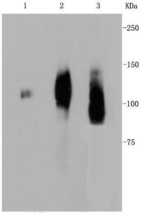
Fig1: Western blot analysis of LAMP2a on different lysates using anti-LAMP2a antibody at 1/1,000 dilution. Positive control: Lane 1: Human placenta Lane 2: JAR Lane 3: Human liverApplication
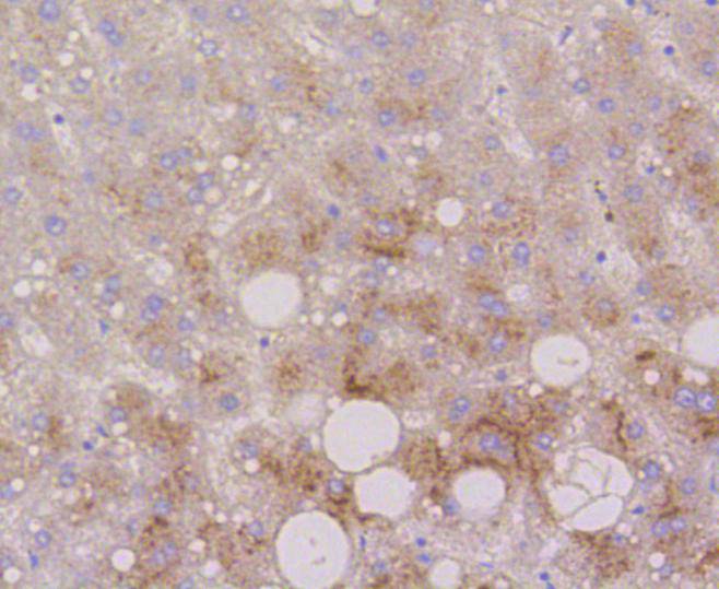
Fig2: Immunohistochemical analysis of paraffin-embedded human liver tissue using anti-LAMP2a antibody. Counter stained with hematoxylin.Application
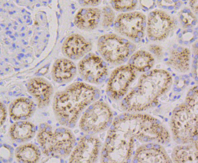
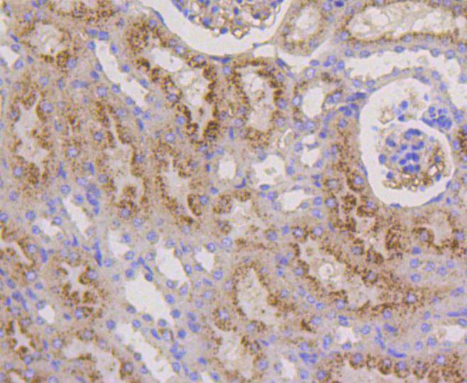
Fig3: Immunohistochemical analysis of paraffin-embedded human kidney tissue using anti-LAMP2a antibody. Counter stained with hematoxylin.Application
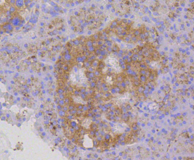
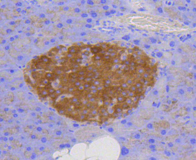
Fig4: Immunohistochemical analysis of paraffin-embedded human pancreas tissue using anti-LAMP2a antibody. Counter stained with hematoxylin.Application
Fig5: Immunohistochemical analysis of paraffin-embedded mouse kidney tissue using anti-LAMP2a antibody. Counter stained with hematoxylin.Application
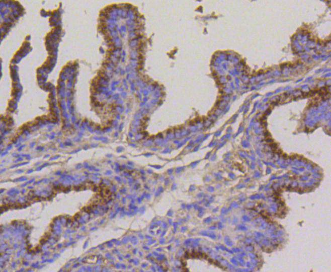
Fig6: Immunohistochemical analysis of paraffin-embedded mouse placenta tissue using anti-LAMP2a antibody. Counter stained with hematoxylin.Application
Fig7: Immunohistochemical analysis of paraffin-embedded mouse brain tissue using anti-LAMP2a antibody. Counter stained with hematoxylin.| Product Name | LAMP2a [SA46-01] |
|---|---|
| Antibody Type | Primary Antibodies |
| Antigen Alias | CD107 antigen-like family member B antibody CD107b antibody LAMP 2 antibody Lamp 2a antibody LAMP-2 antibody LAMP2 antibody LAMP2_HUMAN antibody Lysosome-associated membrane glycoprotein 2 antibody Lysosome-associated membrane protein 2 antibody |
| Product description | Lysosome-associated membrane proteins (LAMP) are glycosylated type I membrane proteins that play a role in the biogenesis of the pigment melanin. LAMP-1 (also designated CD107A) and LAMP-2 (also designated CD107B) are involved in a variety of functions, including cellular adhesion, and are thought to participate in the process of tumor invasion and metastasis. Newly synthesized LAMP-1 and LAMP-2 proteins are sorted at the trans Golgi network and are transported intracellularly via a pathway that is distinct from the clathrin-coated vesicles used for the mannose-6 phosphate receptor. LAMP-1 is expressed on the surface of thrombin-activated but not resting platelets, and it is thought to be involved in the adhesive, prothrombic properties of these cells. Both LAMP-1 and LAMP-2 are involved in maintaining lysosome acidity and protecting the lysosomal membranes from autodigestion, and their expression is increased in patients with lysosomal storage disorders. |
| Immunogen | recombinant protein |
| Clonality | Monoclonal |
|---|---|
| Isotype | IgG |
| Host Species | Recombinant rabbit |
| Tested Applications | WBIHCIP |
| WB:1:1,000-1:5,000 IHC:1:50-1:200 |
|
| Species Reactivity | HumanMouseRat |
| Concentration | 1mg/ml |
| Alternative Names | CD107 antigen-like family member B antibody CD107b antibody LAMP 2 antibody Lamp 2a antibody LAMP-2 antibody LAMP2 antibody LAMP2_HUMAN antibody Lysosome-associated membrane glycoprotein 2 antibody Lysosome-associated membrane protein 2 antibody |
|---|---|
| Molecular Weight(MW) | 120 kDa |
| Cellular Localization | Cell membrane, Endosome membrane, Lysosome membrane |
Application
Fig1: Western blot analysis of LAMP2a on different lysates using anti-LAMP2a antibody at 1/1,000 dilution. Positive control: Lane 1: Human placenta Lane 2: JAR Lane 3: Human liver
Application
Fig2: Immunohistochemical analysis of paraffin-embedded human liver tissue using anti-LAMP2a antibody. Counter stained with hematoxylin.
Application
Fig3: Immunohistochemical analysis of paraffin-embedded human kidney tissue using anti-LAMP2a antibody. Counter stained with hematoxylin.
Application
Fig4: Immunohistochemical analysis of paraffin-embedded human pancreas tissue using anti-LAMP2a antibody. Counter stained with hematoxylin.
Application
Fig5: Immunohistochemical analysis of paraffin-embedded mouse kidney tissue using anti-LAMP2a antibody. Counter stained with hematoxylin.
Application
Fig6: Immunohistochemical analysis of paraffin-embedded mouse placenta tissue using anti-LAMP2a antibody. Counter stained with hematoxylin.
Application
Fig7: Immunohistochemical analysis of paraffin-embedded mouse brain tissue using anti-LAMP2a antibody. Counter stained with hematoxylin.| Positive Control | JAR, human kidney tissue, human liver tissue, human pancreas tissue, mouse placenta tissue, mouse kidney tissue, mouse brain tissue. |
|---|---|
| Application Notes | WB:1:1,000-1:5,000 IHC:1:50-1:200 |
| Form | Liquid |
|---|---|
| Storage Instructions | Store at +4℃ after thawing. Aliquot store at -20℃ or -80℃. Avoid repeated freeze / thaw cycles. |
| Storage Buffer | 1*TBS (pH7.4), 1%BSA, 40%Glycerol. Preservative: 0.05% Sodium Azide. |


