Application
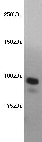
Fig1: Western blot analysis of CD10 on mouse kidney lysate using anti-CD10 antibody at 1/500 dilution.Application
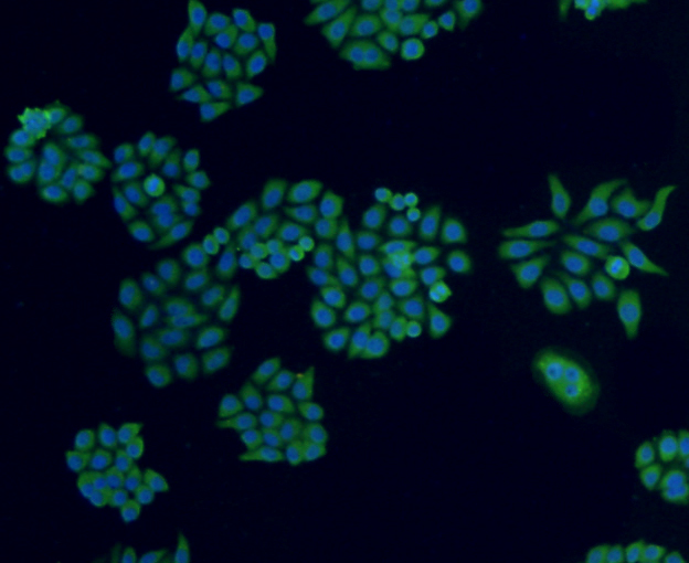
Fig2: ICC staining CD10 in HepG2 cells (green). The nuclear counter stain is DAPI (blue). Cells were fixed in paraformaldehyde, permeabilised with 0.25% Triton X100/PBS.Application
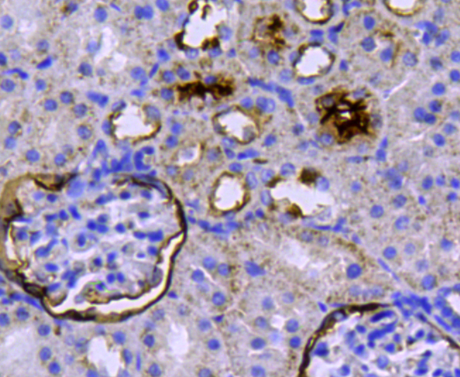
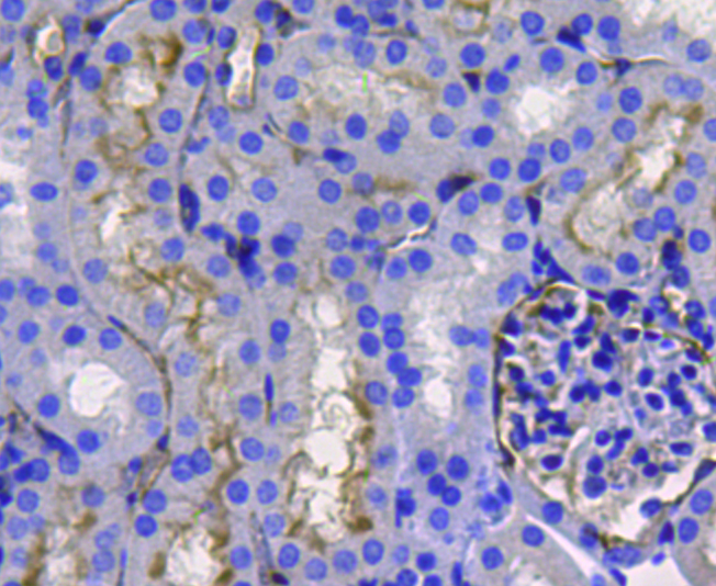
Fig3: Immunohistochemical analysis of paraffin-embedded rat kidney tissue using anti-CD10 antibody. Counter stained with hematoxylin.Application
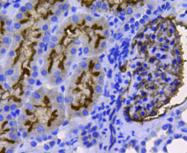
Fig4: Immunohistochemical analysis of paraffin-embedded human kidney tissue using anti-CD10 antibody. Counter stained with hematoxylin.Application
Fig5: Immunohistochemical analysis of paraffin-embedded mouse kidney tissue using anti-CD10 antibody. Counter stained with hematoxylin.Application
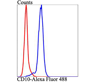
Fig6: Flow cytometric analysis of Jurkat cells with CD10 antibody at 1/100 dilution (blue) compared with an unlabelled control (cells without incubation with primary antibody; red). Alexa Fluor 488-conjugated goat anti-rabbit IgG was used as the secondary antibody.| Product Name | CD10 |
|---|---|
| Antibody Type | Primary Antibodies |
| Antigen Alias | Atriopeptidase antibody CALLA antibody CD10 antibody CD10 antigen antibody Common acute lymphocytic leukemia antigen antibody DKFZp686O16152 antibody EC 3.4.24.11 antibody Enkephalinase antibody EPN antibody Membrane metallo endopeptidase (neutral endopeptidase, enkephalinase) antibody Membrane metallo endopeptidase (neutral endopeptidase, enkephalinase, CALLA, CD10) antibody Membrane metallo endopeptidase antibody Membrane metallo endopeptidase variant 1 antibody Membrane metallo endopeptidase variant 2 antibody Membrane metalloendopeptidase antibody Membrane metalloendopeptidase neutral endopeptidase enkephalinase antibody Membrane metalloendopeptidase neutral endopeptidase enkephalinase CALLA CD10 antibody Membrane metalloendopeptidase variant 1 antibody Membrane metalloendopeptidase variant 2 antibody MGC126681 antibody MGC126707 antibody MME antibody NEP antibody NEP_HUMAN antibody Neprilysin antibody neprilysin-390 antibody neprilysin-411 antibody Neutral endopeptidase 24.11 antibody Neutral endopeptidase antibody Neutral endopeptidase, membrane-associated antibody SFE antibody Skin fibroblast elastase antibody |
| Product description | Thermolysin-like specificity, but is almost confined on acting on polypeptides of up to 30 amino acids. Biologically important in the destruction of opioid peptides such as Met- and Leu-enkephalins by cleavage of a Gly-Phe bond. Able to cleave angiotensin-1, angiotensin-2 and angiotensin 1-9. Involved in the degradation of atrial natriuretic factor (ANF). Displays UV-inducible elastase activity toward skin preelastic and elastic fibers. |
| Immunogen | This antibody is produced by immunizing rabbits with a synthetic peptide (KLH-coupled) corresponding to a region of CD-10. |
| Clonality | Polyclonal |
|---|---|
| Isotype | IgG |
| Host Species | Rabbit |
| Tested Applications | WBICCIHCFC |
| WB:1:500 ICC:1:50-1:200 IHC:1:50-1:200 FC:1:50-1:100 |
|
| Species Reactivity | HumanMouseRat |
| Concentration | 1mg/ml |
| Alternative Names | Atriopeptidase antibody
CALLA antibody
CD10 antibody
CD10 antigen antibody
Common acute lymphocytic leukemia antigen antibody
DKFZp686O16152 antibody
EC 3.4.24.11 antibody
Enkephalinase antibody
EPN antibody
Membrane metallo endopeptidase (neutral endopeptidase enkephalinase) antibody Membrane metallo endopeptidase (neutral endopeptidase enkephalinase CALLA CD10) antibody Membrane metallo endopeptidase antibody Membrane metallo endopeptidase variant 1 antibody Membrane metallo endopeptidase variant 2 antibody Membrane metalloendopeptidase antibody Membrane metalloendopeptidase neutral endopeptidase enkephalinase antibody Membrane metalloendopeptidase neutral endopeptidase enkephalinase CALLA CD10 antibody Membrane metalloendopeptidase variant 1 antibody Membrane metalloendopeptidase variant 2 antibody MGC126681 antibody MGC126707 antibody MME antibody NEP antibody NEP_HUMAN antibody Neprilysin antibody neprilysin-390 antibody neprilysin-411 antibody Neutral endopeptidase 24.11 antibody Neutral endopeptidase antibody Neutral endopeptidase membrane-associated antibody SFE antibody Skin fibroblast elastase antibody |
|---|---|
| Molecular Weight(MW) | 85kDa |
| Cellular Localization | Cell membrane. Membrane. |
Application
Fig1: Western blot analysis of CD10 on mouse kidney lysate using anti-CD10 antibody at 1/500 dilution.
Application
Fig2: ICC staining CD10 in HepG2 cells (green). The nuclear counter stain is DAPI (blue). Cells were fixed in paraformaldehyde, permeabilised with 0.25% Triton X100/PBS.
Application
Fig3: Immunohistochemical analysis of paraffin-embedded rat kidney tissue using anti-CD10 antibody. Counter stained with hematoxylin.
Application
Fig4: Immunohistochemical analysis of paraffin-embedded human kidney tissue using anti-CD10 antibody. Counter stained with hematoxylin.
Application
Fig5: Immunohistochemical analysis of paraffin-embedded mouse kidney tissue using anti-CD10 antibody. Counter stained with hematoxylin.
Application
Fig6: Flow cytometric analysis of Jurkat cells with CD10 antibody at 1/100 dilution (blue) compared with an unlabelled control (cells without incubation with primary antibody; red). Alexa Fluor 488-conjugated goat anti-rabbit IgG was used as the secondary antibody.| Positive Control | Human kidney tissue, mouse kidney tissue, rat kidney tissue, Jurkat, HepG2. |
|---|---|
| Application Notes | WB:1:500 ICC:1:50-1:200 IHC:1:50-1:200 FC:1:50-1:100 |
| Form | Liquid |
|---|---|
| Storage Instructions | Store at +4℃ after thawing. Aliquot store at -20℃. Avoid repeated freeze / thaw cycles. |
| Storage Buffer | 1*TBS (pH7.4), 1%BSA, 25%Glycerol. Preservative: 0.05% Sodium Azide. |


