Application
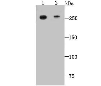
Fig1: Western blot analysis of Dopey-2 on mouse heart (1) and human heart (2) tissue lysate using anti-Dopey-2 antibody at 1/500 dilution.Application
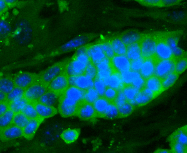
Fig2: ICC staining Dopey-2 in D3 cells (green). The nuclear counter stain is DAPI (blue). Cells were fixed in paraformaldehyde, permeabilised with 0.25% Triton X100/PBS.Application
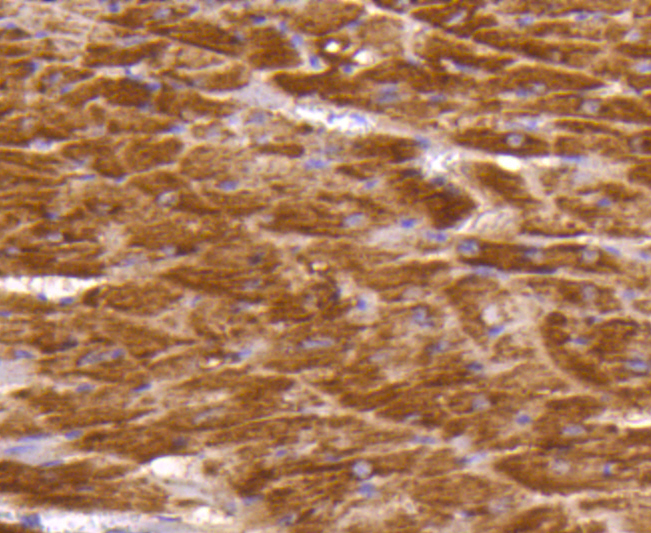
Fig3: Immunohistochemical analysis of formalin-fixed, paraffin-embedded mouse heart tissue labeling Dopey-2. Counterstained with Hematoxylin.Application
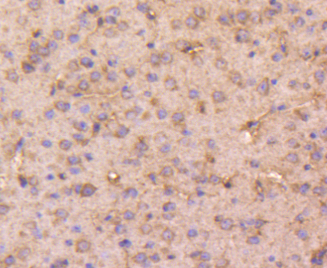
Fig4: Immunohistochemical analysis of formalin-fixed, paraffin-embedded mouse brain tissue labeling Dopey-2. Counterstained with Hematoxylin.Application
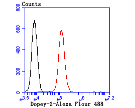
Fig5: Flow cytometric analysis of N2A cells with Dopey-2 antibody at 1/100 dilution (red) compared with an unlabelled control (cells without incubation with primary antibody; black).Alexa Fluor 488-conjugated goat anti-rabbit IgG was used as the secondary antibody.| Product Name | DOPEY2 |
|---|---|
| Antibody Type | Primary Antibodies |
| Antigen Alias | Dipeptidase 2 antibody Dipeptidase2 antibody DPEP 2 antibody MBD 2 antibody MBD2 antibody |
| Product description | Dopey-2, a 2,298 amino acid protein, belongs to the dopey family, that is overexpressed in lymphoblasts from Down syndrome patients and Abundantly expressed in developing central nervous system, with highest levels in cerebellum and lowest in telencephalon. Multiple isoforms of Dopey-2 exist due to alternative splicing events. Dopey-2 is thought to play a role in protein trafficking between early endosomes and the late. |
| Immunogen | Peptide |
| Clonality | Polyclonal |
|---|---|
| Isotype | IgG |
| Host Species | Rabbit |
| Tested Applications | WBICCIHCFC |
| WB:1:500-1:1,000 IHC:1:50-1:200 ICC:1:50-1:200 FC:1:50-1:100 |
|
| Species Reactivity | HumanMouse |
| Concentration | 1mg/ml |
| Alternative Names | Dipeptidase 2 antibody Dipeptidase2 antibody DPEP 2 antibody MBD 2 antibody MBD2 antibody |
|---|---|
| Molecular Weight(MW) | 260kDa |
| Cellular Localization | Cytosol. Endosome. |
Application
Fig1: Western blot analysis of Dopey-2 on mouse heart (1) and human heart (2) tissue lysate using anti-Dopey-2 antibody at 1/500 dilution.
Application
Fig2: ICC staining Dopey-2 in D3 cells (green). The nuclear counter stain is DAPI (blue). Cells were fixed in paraformaldehyde, permeabilised with 0.25% Triton X100/PBS.
Application
Fig3: Immunohistochemical analysis of formalin-fixed, paraffin-embedded mouse heart tissue labeling Dopey-2. Counterstained with Hematoxylin.
Application
Fig4: Immunohistochemical analysis of formalin-fixed, paraffin-embedded mouse brain tissue labeling Dopey-2. Counterstained with Hematoxylin.
Application
Fig5: Flow cytometric analysis of N2A cells with Dopey-2 antibody at 1/100 dilution (red) compared with an unlabelled control (cells without incubation with primary antibody; black).Alexa Fluor 488-conjugated goat anti-rabbit IgG was used as the secondary antibody.| Positive Control | Mouse heart tissue lysate, human heart tissue lysate, D3 cells, N2A cells, mouse heart tissue, mouse brain tissue. |
|---|---|
| Application Notes | WB:1:500-1:1,000 IHC:1:50-1:200 ICC:1:50-1:200 FC:1:50-1:100 |
| Form | Liquid |
|---|---|
| Storage Instructions | Store at +4℃ after thawing. Aliquot store at -20℃ or -80℃. Avoid repeated freeze / thaw cycles. |
| Storage Buffer | 1*TBS (pH7.4), 1%BSA, 40%Glycerol. Preservative: 0.05% Sodium Azide. |


