WB
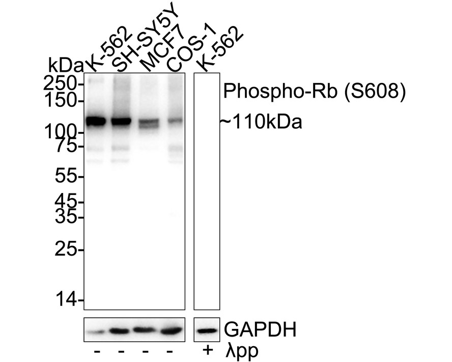
Western blot analysis of Phospho-Rb (S608) on different lysates with Rabbit anti-Phospho-Rb (S608) antibody at 1/5,000 dilution. Lane 1: K-562 cell lysate (no heat), Lane 2: SH-SY5Y cell lysate (no heat), Lane 3: MCF7 cell lysate (no heat), Lane 4: COS-1 cell lysate (no heat), Lane 5: K-562 cell lysate (no heat), the membrane treated with λpp for 1 hour, Notice: no heat means the lysate is not boiled. Lysates/proteins at 20 µg/Lane. Exposure time: 12 seconds; 4-20% SDS-PAGE gel. Proteins were transferred to a PVDF membrane and blocked with 5% NFDM/TBST for 1 hour at room temperature. The primary antibody at 1/5,000 dilution was used in 5% NFDM/TBST at 4℃ overnight. Goat Anti-Rabbit IgG - HRP Secondary Antibody at 1/50,000 dilution was used for 1 hour at room temperature.ICC/IF
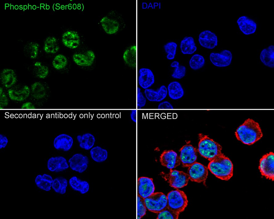
Immunocytochemistry analysis of K-562 cells labeling Phospho-Rb (S608) with Rabbit anti-Phospho-Rb (S608) antibody at 1/100 dilution. Cells were fixed in 4% paraformaldehyde for 15 minutes at room temperature, permeabilized with 0.1% Triton X-100 in PBS for 15 minutes at room temperature, then blocked with 1% BSA in 10% negative goat serum for 1 hour at room temperature. Cells were then incubated with Rabbit anti-Phospho-Rb (S608) antibody at 1/100 dilution in 1% BSA in PBST overnight at 4 ℃. Goat Anti-Rabbit IgG H&L (488) was used as the secondary antibody at 1/1,000 dilution. PBS instead of the primary antibody was used as the secondary antibody only control. Nuclear DNA was labelled in blue with DAPI. Beta tubulin (red) was stained at 1/100 dilution overnight at +4℃. Goat Anti-Mouse IgG H&L (594) was used as the secondary antibody at 1/1,000 dilution.| Product Name | Phospho-Rb (S608) Recombinant Rabbit Monoclonal Antibody |
|---|---|
| Antibody Type | Primary Antibodies |
| Product description | The retinoblastoma protein (protein name abbreviated Rb or pRb; gene name abbreviated Rb, RB or RB1) is a tumor suppressor protein that is dysfunctional in several major cancers. One function of pRb is to prevent excessive cell growth by inhibiting cell cycle progression until a cell is ready to divide. When the cell is ready to divide, pRb is phosphorylated, inactivating it, and the cell cycle is allowed to progress. It is also a recruiter of several chromatin remodeling enzymes such as methylases and acetylases. pRb belongs to the pocket protein family, whose members have a pocket for the functional binding of other proteins. Should an oncogenic protein, such as those produced by cells infected by high-risk types of human papillomavirus, bind and inactivate pRb, this can lead to cancer. The RB gene may have been responsible for the evolution of multicellularity in several lineages of life including animals. |
| Immunogen | Synthetic phospho-peptide corresponding to residues surrounding Ser608 of Human Rb. |
| Modification | p-S608 |
| Clonality | Monoclonal |
|---|---|
| Isotype | IgG |
| Host Species | Recombinant rabbit |
| Tested Applications | ICC/IFWB |
| WB:1:5000 ICC/IF:1:100 |
|
| Species Reactivity | HumanMonkey |
| Concentration | 1mg/ml |
| Purification | Protein A |
| Alternative Names | OSRC antibody Osteosarcoma antibody p105-Rb antibody P105RB antibody PP105 antibody pp110 antibody PPP1R130 antibody pRb antibody Prepro retinoblastoma associated protein antibody Protein phosphatase 1 regulatory subunit 130 antibody Rb antibody RB_HUMAN antibody RB1 antibody RB1 gene antibody Retinoblastoma 1 antibody Retinoblastoma suspectibility protein antibody Retinoblastoma-associated protein antibody |
|---|---|
| Molecular Weight(MW) | 106 kDa(Observed band size: 110 kDa) |
| Cellular Localization | Nucleus, Cytoplasm. |
WB
Western blot analysis of Phospho-Rb (S608) on different lysates with Rabbit anti-Phospho-Rb (S608) antibody at 1/5,000 dilution. Lane 1: K-562 cell lysate (no heat), Lane 2: SH-SY5Y cell lysate (no heat), Lane 3: MCF7 cell lysate (no heat), Lane 4: COS-1 cell lysate (no heat), Lane 5: K-562 cell lysate (no heat), the membrane treated with λpp for 1 hour, Notice: no heat means the lysate is not boiled. Lysates/proteins at 20 µg/Lane. Exposure time: 12 seconds; 4-20% SDS-PAGE gel. Proteins were transferred to a PVDF membrane and blocked with 5% NFDM/TBST for 1 hour at room temperature. The primary antibody at 1/5,000 dilution was used in 5% NFDM/TBST at 4℃ overnight. Goat Anti-Rabbit IgG - HRP Secondary Antibody at 1/50,000 dilution was used for 1 hour at room temperature.
ICC/IF
Immunocytochemistry analysis of K-562 cells labeling Phospho-Rb (S608) with Rabbit anti-Phospho-Rb (S608) antibody at 1/100 dilution. Cells were fixed in 4% paraformaldehyde for 15 minutes at room temperature, permeabilized with 0.1% Triton X-100 in PBS for 15 minutes at room temperature, then blocked with 1% BSA in 10% negative goat serum for 1 hour at room temperature. Cells were then incubated with Rabbit anti-Phospho-Rb (S608) antibody at 1/100 dilution in 1% BSA in PBST overnight at 4 ℃. Goat Anti-Rabbit IgG H&L (488) was used as the secondary antibody at 1/1,000 dilution. PBS instead of the primary antibody was used as the secondary antibody only control. Nuclear DNA was labelled in blue with DAPI. Beta tubulin (red) was stained at 1/100 dilution overnight at +4℃. Goat Anti-Mouse IgG H&L (594) was used as the secondary antibody at 1/1,000 dilution.| Application Notes | WB:1:5000 ICC/IF:1:100 |
|---|
| Form | Liquid |
|---|---|
| Storage Instructions | Store at +4℃ after thawing. Aliquot store at -20℃ or -80℃. Avoid repeated freeze / thaw cycles. |
| Storage Buffer | PBS (pH7.4), 0.1% BSA, 40% Glycerol. Preservative: 0.05% Sodium Azide. |


