Application
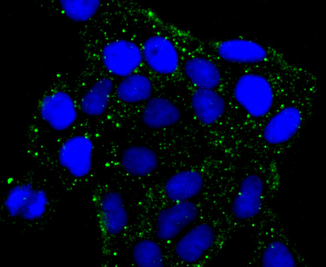
Fig1: ICC staining PKA 2 beta (regulatory subunit) in Hela cells (green). The nuclear counter stain is DAPI (blue). Cells were fixed in paraformaldehyde, permeabilised with 0.25% Triton X100/PBS.Application
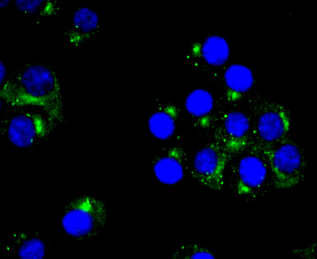
Fig2: ICC staining PKA 2 beta (regulatory subunit) in N2A cells (green). The nuclear counter stain is DAPI (blue). Cells were fixed in paraformaldehyde, permeabilised with 0.25% Triton X100/PBS.Application
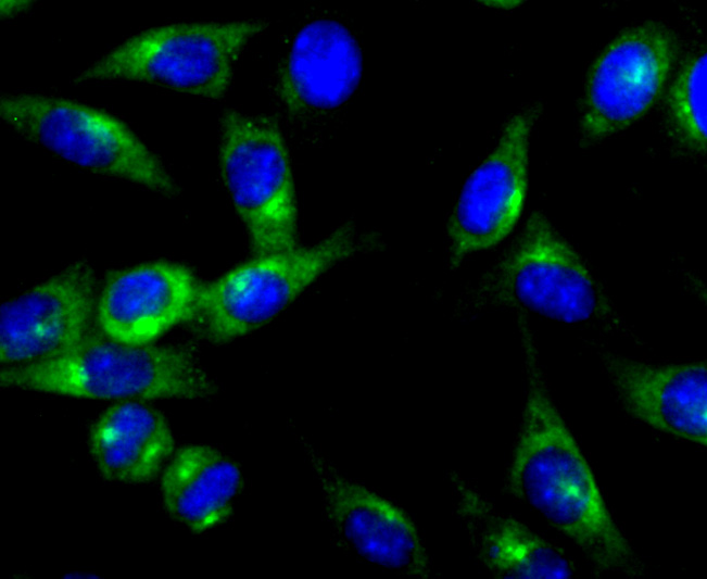
Fig3: ICC staining PKA 2 beta (regulatory subunit) in SH-SY5Y cells (green). The nuclear counter stain is DAPI (blue). Cells were fixed in paraformaldehyde, permeabilised with 0.25% Triton X100/PBS.Application
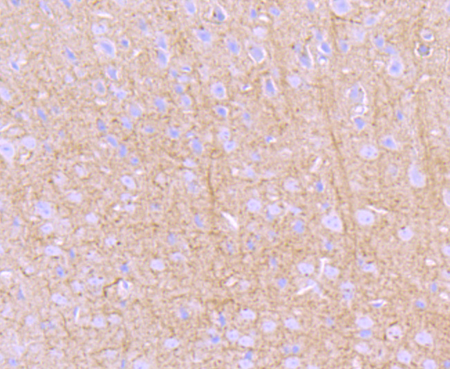
Fig4: Immunohistochemical analysis of paraffin-embedded mouse brain tissue using anti- PKA 2 beta (regulatory subunit) antibody. Counter stained with hematoxylin.Application
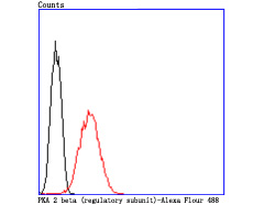
Fig5: Flow cytometric analysis of N2A cells with PKA 2 beta (regulatory subunit) antibody at 1/50 dilution (red) compared with an unlabelled control (cells without incubation with primary antibody; black). Alexa Fluor 488-conjugated goat anti rabbit IgG was used as the secondary antibody.| Product Name | PKA 2 beta (regulatory subunit) [JA11-69] |
|---|---|
| Antibody Type | Primary Antibodies |
| Antigen Alias | AI451071 antibody AW061005 antibody cAMP dependent protein kinase type II beta regulatory chain antibody cAMP dependent protein kinase type II beta regulatory subunit antibody cAMP-dependent protein kinase type II-beta regulatory subunit antibody H RG363E19.2 antibody KAP3_HUMAN antibody MGC116401 antibody Pkarb2 antibody PRKAR 2 antibody PRKAR2 antibody PRKAR2B antibody Protein kinase cAMP dependent regulatory type II beta antibody RATDNA antibody RII beta antibody RII(beta) antibody RIIbeta antibody WUGSC:H RG363E19.2 antibody |
| Product description | The second messenger cyclic AMP mediates diverse cellular responses to external signals such as proliferation, ion transport, regulation of metabolism and gene transcription by activation of the cAMP-dependent protein kinase (cAPK or PKA). Activation of PKA occurs when cAMP binds to the two regulatory subunits of the tetrameric PKA holoenzyme resulting in release of active catalytic subunits. One of several regulatory subunits, p-PKA IIβ reg (cAMP-dependent protein kinase type II-beta regulatory subunit), also known as PRKAR2B, is a 418 amino acid protein that is phosphorylated by the activated catalytic chain. p-PKA IIβ reg knockout mice exhibit diminished white adipose tissue and were protected against diet-induced obesity and fatty livers,as well as markedly reduced leptin mRNA. Also playing a role in the immune response, p-PKA IIβ reg suppresses CREB transcriptional activity and down-regulates IL-2 production in T-lymphocytes. |
| Immunogen | recombinant protein |
| Clonality | Monoclonal |
|---|---|
| Isotype | IgG |
| Host Species | Recombinant rabbit |
| Tested Applications | WBIPICC/IFIHCFC |
| WB:1:500-1:1,000 ICC:1:50-1:200 IHC:1:50-1:200 FC:1:50-1:100 |
|
| Species Reactivity | HumanMouseRat |
| Concentration | 1mg/ml |
| Alternative Names | AI451071 antibody AW061005 antibody cAMP dependent protein kinase type II beta regulatory chain antibody cAMP dependent protein kinase type II beta regulatory subunit antibody cAMP-dependent protein kinase type II-beta regulatory subunit antibody H RG363E19.2 antibody KAP3_HUMAN antibody MGC116401 antibody Pkarb2 antibody PRKAR 2 antibody PRKAR2 antibody PRKAR2B antibody Protein kinase cAMP dependent regulatory type II beta antibody RATDNA antibody RII beta antibody RII(beta) antibody RIIbeta antibody WUGSC:H RG363E19.2 antibody |
|---|---|
| Molecular Weight(MW) | 46 kDa |
| Cellular Localization | Cytoplasm. Cell membrane. |
Application
Fig1: ICC staining PKA 2 beta (regulatory subunit) in Hela cells (green). The nuclear counter stain is DAPI (blue). Cells were fixed in paraformaldehyde, permeabilised with 0.25% Triton X100/PBS.
Application
Fig2: ICC staining PKA 2 beta (regulatory subunit) in N2A cells (green). The nuclear counter stain is DAPI (blue). Cells were fixed in paraformaldehyde, permeabilised with 0.25% Triton X100/PBS.
Application
Fig3: ICC staining PKA 2 beta (regulatory subunit) in SH-SY5Y cells (green). The nuclear counter stain is DAPI (blue). Cells were fixed in paraformaldehyde, permeabilised with 0.25% Triton X100/PBS.
Application
Fig4: Immunohistochemical analysis of paraffin-embedded mouse brain tissue using anti- PKA 2 beta (regulatory subunit) antibody. Counter stained with hematoxylin.
Application
Fig5: Flow cytometric analysis of N2A cells with PKA 2 beta (regulatory subunit) antibody at 1/50 dilution (red) compared with an unlabelled control (cells without incubation with primary antibody; black). Alexa Fluor 488-conjugated goat anti rabbit IgG was used as the secondary antibody.| Positive Control | Hela, N2A, SH-SY5Y, mouse brain tissue. |
|---|---|
| Application Notes | WB:1:500-1:1,000 ICC:1:50-1:200 IHC:1:50-1:200 FC:1:50-1:100 |
| Form | Liquid |
|---|---|
| Storage Instructions | Store at +4℃ after thawing. Aliquot store at -20℃ or -80℃. Avoid repeated freeze / thaw cycles. |
| Storage Buffer | 1*TBS (pH7.4), 1%BSA, 40%Glycerol. Preservative: 0.05% Sodium Azide. |


