Application
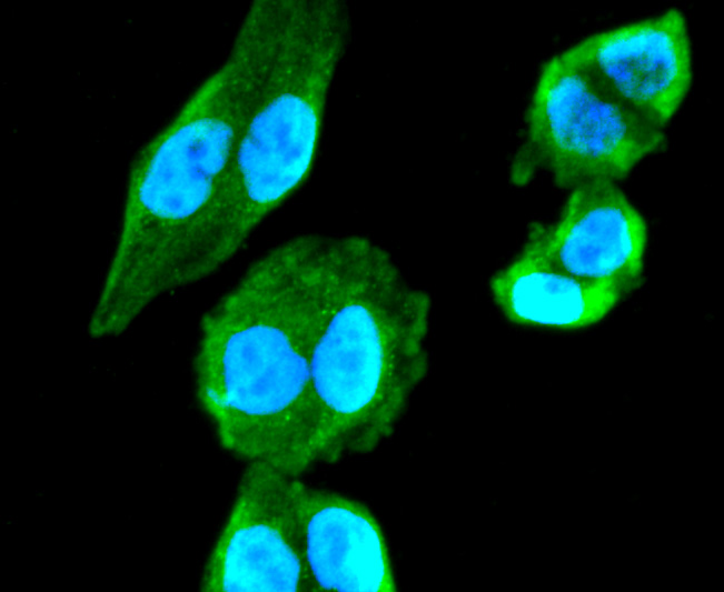
Fig1: ICC staining Cho Line Acetyltransferase in Hela cells (green). The nuclear counter stain is DAPI (blue). Cells were fixed in paraformaldehyde, permeabilised with 0.25% Triton X100/PBS.Application
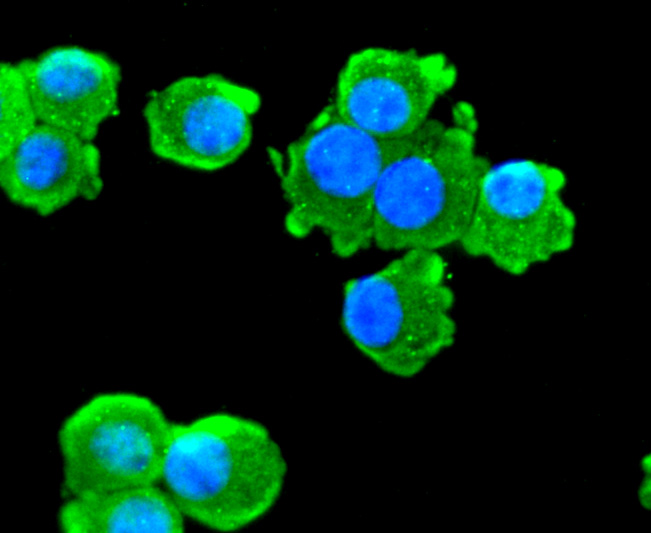
Fig2: ICC staining Choline Acetyltransferase in N2A cells (green). The nuclear counter stain is DAPI (blue). Cells were fixed in paraformaldehyde, permeabilised with 0.25% Triton X100/PBS.Application
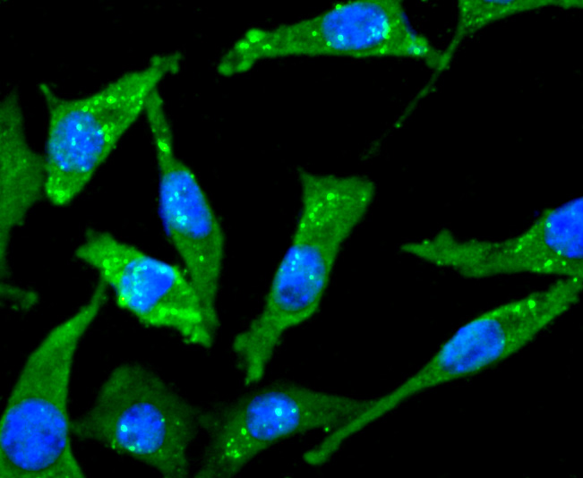
Fig3: ICC staining Choline Acetyltransferase in SH-SY5Y cells (green). The nuclear counter stain is DAPI (blue). Cells were fixed in paraformaldehyde, permeabilised with 0.25% Triton X100/PBS.Application
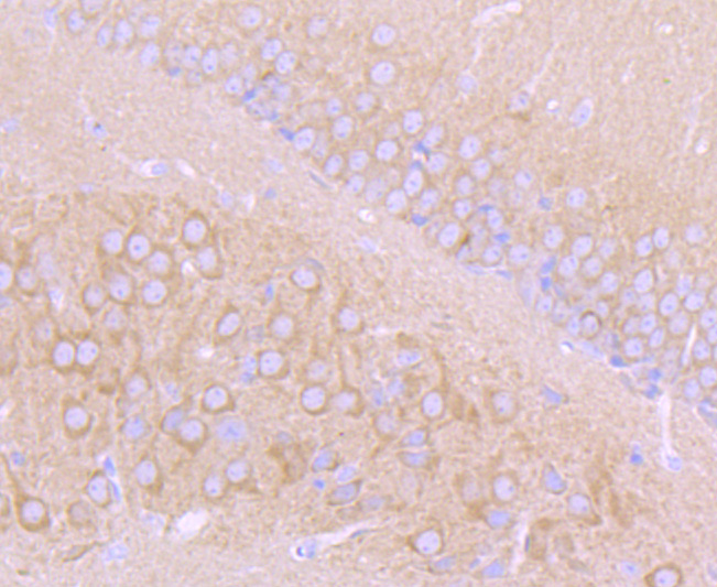
Fig4: Immunohistochemical analysis of paraffin-embedded mouse brain tissue using anti- Choline Acetyltransferase antibody. Counter stained with hematoxylin.Application
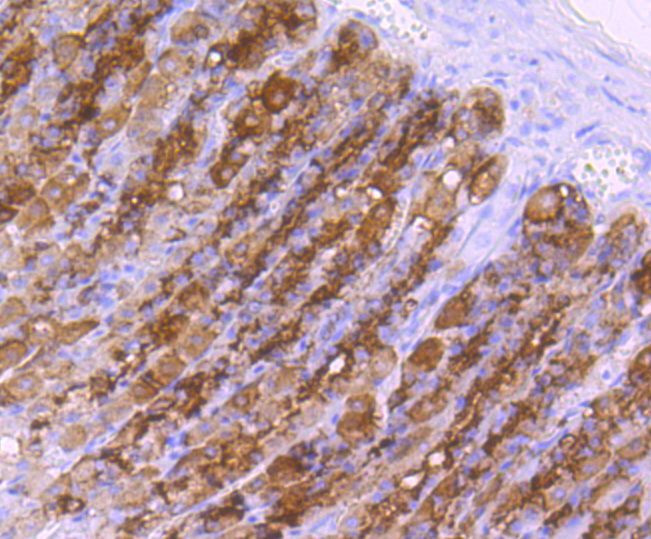
Fig5: Immunohistochemical analysis of paraffin-embedded rat stomach tissue using anti- Choline Acetyltransferase antibody. Counter stained with hematoxylin.Application
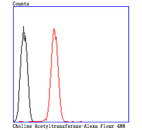
Fig6: Flow cytometric analysis of N2A cells with Choline Acetyltransferase antibody at 1/50 dilution (red) compared with an unlabelled control (cells without incubation with primary antibody; black). Alexa Fluor 488-conjugated goat anti rabbit IgG was used as the secondary antibody.| Product Name | Choline Acetyltransferase [JA67-11] |
|---|---|
| Antibody Type | Primary Antibodies |
| Antigen Alias | Acetyl CoA choline O acetyltransferase antibody Acetyl CoA:choline O acetyltransferase antibody ChAT antibody CHOACTase antibody Choline acetylase antibody choline acetyltransferase antibody Choline O acetyltransferase antibody Choline O-acetyltransferase antibody CLAT_HUMAN antibody CMS1A antibody CMS1A2 antibody EC 2.3.1.6 antibody OTTHUMP00000019583 antibody OTTHUMP00000019584 antibody |
| Product description | Choline acetyltransferase (also designated choactase, choline O-acetyltransferase) synthesizes acetylcholine in cholinergic neurons. Multiple choactase mRNAs with different 5'-noncoding regions are expressed as R-, N1, N2-, S- and M-types. N1-, N2- and R-type mRNAs produce a single short enzyme, while M-type mRNA produces both long and short enzymes. The long enzyme is targeted to the nuclei of cells, whereas the short protein is found in cytoplasm. A novel NFkB binding site is located within the nerve growth factor-responsive enhancer element that is recognized by the NFkB protein p49, but not p65 or p50. Decreased choactase expression and increased NFkB activity are associated with aging and Alzheimer's disease, indicating that p49 is a negative regulator of choactase expression and suggesting a possible mechanism for aging-associated declines in cholinergic function. Phosphorylation of choactase has been shown to enhance choactase catalytic activity. Specifically, Serine 440 is found to be the phosphorylation site in a recombinant human short choactase by protein kinase C and is involved in regulation of the enzyme catalytic activity and binding to subcellular membranes. |
| Immunogen | recombinant protein |
| Clonality | Monoclonal |
|---|---|
| Isotype | IgG |
| Host Species | Recombinant rabbit |
| Tested Applications | WBIPICC/IFIHCFC |
| WB:1:500-1:1,000 ICC:1:50-1:200 IHC:1:50-1:200 FC:1:50-1:100 IP:1:10-1:50 |
|
| Species Reactivity | HumanMouseRat |
| Concentration | 1mg/ml |
| Alternative Names | Acetyl CoA choline O acetyltransferase antibody Acetyl CoA:choline O acetyltransferase antibody ChAT antibody CHOACTase antibody Choline acetylase antibody choline acetyltransferase antibody Choline O acetyltransferase antibody Choline O-acetyltransferase antibody CLAT_HUMAN antibody CMS1A antibody CMS1A2 antibody EC 2.3.1.6 antibody OTTHUMP00000019583 antibody OTTHUMP00000019584 antibody |
|---|---|
| Molecular Weight(MW) | 82 kDa |
| Cellular Localization | Cytoplasm. Nucleus. |
Application
Fig1: ICC staining Cho Line Acetyltransferase in Hela cells (green). The nuclear counter stain is DAPI (blue). Cells were fixed in paraformaldehyde, permeabilised with 0.25% Triton X100/PBS.
Application
Fig2: ICC staining Choline Acetyltransferase in N2A cells (green). The nuclear counter stain is DAPI (blue). Cells were fixed in paraformaldehyde, permeabilised with 0.25% Triton X100/PBS.
Application
Fig3: ICC staining Choline Acetyltransferase in SH-SY5Y cells (green). The nuclear counter stain is DAPI (blue). Cells were fixed in paraformaldehyde, permeabilised with 0.25% Triton X100/PBS.
Application
Fig4: Immunohistochemical analysis of paraffin-embedded mouse brain tissue using anti- Choline Acetyltransferase antibody. Counter stained with hematoxylin.
Application
Fig5: Immunohistochemical analysis of paraffin-embedded rat stomach tissue using anti- Choline Acetyltransferase antibody. Counter stained with hematoxylin.
Application
Fig6: Flow cytometric analysis of N2A cells with Choline Acetyltransferase antibody at 1/50 dilution (red) compared with an unlabelled control (cells without incubation with primary antibody; black). Alexa Fluor 488-conjugated goat anti rabbit IgG was used as the secondary antibody.| Positive Control | Hela, N2A, SH-SY5Y, rat stomach tissue, mouse brain tissue. |
|---|---|
| Application Notes | WB:1:500-1:1,000 ICC:1:50-1:200 IHC:1:50-1:200 FC:1:50-1:100 IP:1:10-1:50 |
| Form | Liquid |
|---|---|
| Storage Instructions | Store at +4℃ after thawing. Aliquot store at -20℃ or -80℃. Avoid repeated freeze / thaw cycles. |
| Storage Buffer | 1*TBS (pH7.4), 1%BSA, 40%Glycerol. Preservative: 0.05% Sodium Azide. |


