Application
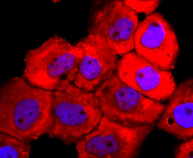
Fig1: ICC staining Tyrosinase in A431 cells (red). The nuclear counter stain is DAPI (blue). Cells were fixed in paraformaldehyde, permeabilised with 0.25% Triton X100/PBS.Application
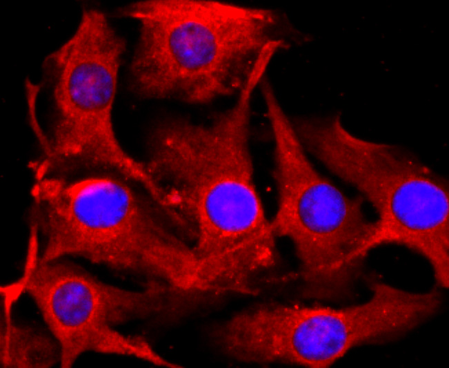
Fig2: ICC staining Tyrosinase in B16F1 cells (red). The nuclear counter stain is DAPI (blue). Cells were fixed in paraformaldehyde, permeabilised with 0.25% Triton X100/PBS.Application
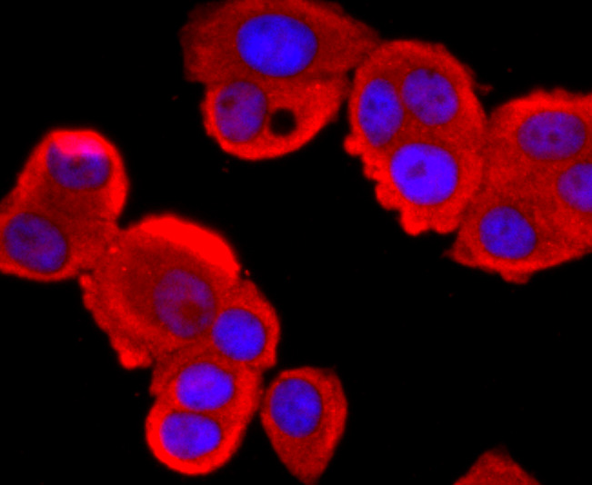
Fig3: ICC staining Tyrosinase in MCF-7 cells (red). The nuclear counter stain is DAPI (blue). Cells were fixed in paraformaldehyde, permeabilised with 0.25% Triton X100/PBS.Application
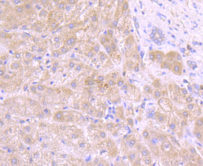
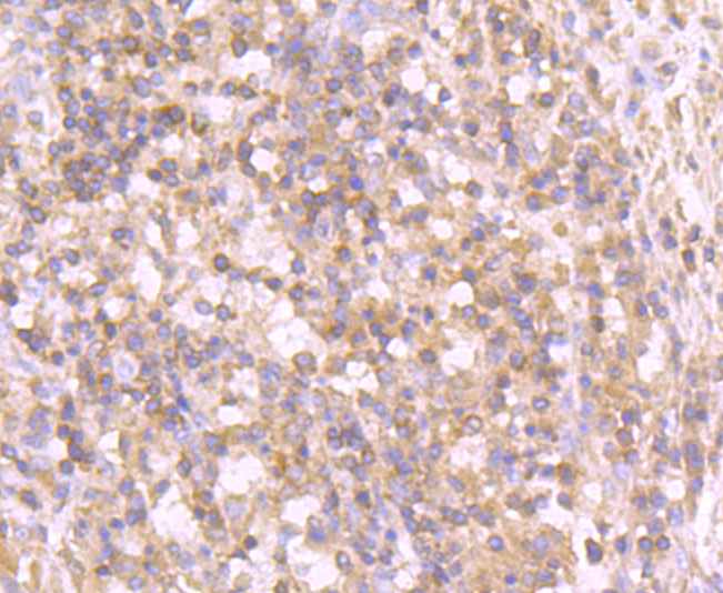
Fig4: Immunohistochemical analysis of paraffin-embedded human liver tissue using anti-Tyrosinase antibody. Counter stained with hematoxylin.Application
Fig5: Immunohistochemical analysis of paraffin-embedded human stomach cancer tissue using anti-Tyrosinase antibody. Counter stained with hematoxylin.| Product Name | Tyrosinase [JA52-11] |
|---|---|
| Antibody Type | Primary Antibodies |
| Antigen Alias | ATN antibody CMM8 antibody LB24 AB antibody LB24-AB antibody Monophenol monooxygenase antibody OCA1 antibody OCA1A antibody OCAIA antibody Oculocutaneous albinism IA antibody SHEP3 antibody SK29 AB antibody SK29-AB antibody Tumor rejection antigen AB antibody TYR antibody TYRO_HUMAN antibody tyrosinase (oculocutaneous albinism IA) antibody Tyrosinase antibody |
| Product description | This is a copper-containing oxidase that functions in the formation of pigments such as melanins and other polyphenolic compounds. Catalyzes the initial and rate limiting step in the cascade of reactions leading to melanin production from tyrosine. In addition to hydroxylating tyrosine to DOPA (3,4-dihydroxyphenylalanine), also catalyzes the oxidation of DOPA to DOPA-quinone, and possibly the oxidation of DHI (5,6-dihydroxyindole) to indole-5,6 quinone. Defects in TYR are the cause of albinism oculocutaneous type 1B (OCA1B) [MIM:606952]; also known as albinism yellow mutant type. An autosomal recessive disorder in which the biosynthesis of melanin pigment is reduced in skin, hair, and eyes. It is characterized by partial lack of tyrosinase activity. |
| Immunogen | recombinant protein |
| Clonality | Monoclonal |
|---|---|
| Isotype | IgG |
| Host Species | Recombinant rabbit |
| Tested Applications | WBICC/IFIHC |
| WB:1:500-1:1,000 ICC:1:100-1:500 IHC:1:50-1:200 |
|
| Species Reactivity | Human |
| Concentration | 1mg/ml |
| Alternative Names | ATN antibody CMM8 antibody LB24 AB antibody LB24-AB antibody Monophenol monooxygenase antibody OCA1 antibody OCA1A antibody OCAIA antibody Oculocutaneous albinism IA antibody SHEP3 antibody SK29 AB antibody SK29-AB antibody Tumor rejection antigen AB antibody TYR antibody TYRO_HUMAN antibody tyrosinase (oculocutaneous albinism IA) antibody Tyrosinase antibody |
|---|---|
| Molecular Weight(MW) | 80 kDa |
| Cellular Localization | Melanosome membrane. |
| SwissProt ID | P14679 |
|---|
Application
Fig1: ICC staining Tyrosinase in A431 cells (red). The nuclear counter stain is DAPI (blue). Cells were fixed in paraformaldehyde, permeabilised with 0.25% Triton X100/PBS.
Application
Fig2: ICC staining Tyrosinase in B16F1 cells (red). The nuclear counter stain is DAPI (blue). Cells were fixed in paraformaldehyde, permeabilised with 0.25% Triton X100/PBS.
Application
Fig3: ICC staining Tyrosinase in MCF-7 cells (red). The nuclear counter stain is DAPI (blue). Cells were fixed in paraformaldehyde, permeabilised with 0.25% Triton X100/PBS.
Application
Fig4: Immunohistochemical analysis of paraffin-embedded human liver tissue using anti-Tyrosinase antibody. Counter stained with hematoxylin.
Application
Fig5: Immunohistochemical analysis of paraffin-embedded human stomach cancer tissue using anti-Tyrosinase antibody. Counter stained with hematoxylin.| Positive Control | A431, B16F1, MCF-7, human liver tissue, human stomach cancer tissue. |
|---|---|
| Application Notes | WB:1:500-1:1,000 ICC:1:100-1:500 IHC:1:50-1:200 |
| Form | Liquid |
|---|---|
| Storage Instructions | Store at +4℃ after thawing. Aliquot store at -20℃ or -80℃. Avoid repeated freeze / thaw cycles. |
| Storage Buffer | 1*TBS (pH7.4), 1%BSA, 40%Glycerol. Preservative: 0.05% Sodium Azide. |


