Application
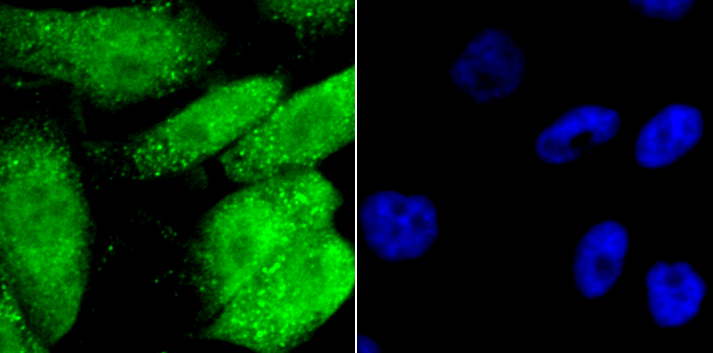
Fig1: ICC staining GATA4 in PC-3M cells (Green). The nuclear counter stain is DAPI (blue). Cells were fixed in paraformaldehyde, permeabilised with 0.25% Triton X100/PBS.Application
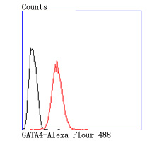
Fig2: Flow cytometric analysis of Hela cells with GATA4 antibody at 1/50 dilution (red) compared with an unlabelled control (cells without incubation with primary antibody; black). Alexa Fluor 488-conjugated goat anti rabbit IgG was used as the secondary antibody.| Product Name | GATA4 [JA11-41] |
|---|---|
| Antibody Type | Primary Antibodies |
| Antigen Alias | ASD2 antibody GATA 4 antibody GATA binding protein 4 antibody GATA-binding factor 4 antibody gata4 antibody GATA4_HUMAN antibody MGC126629 antibody Transcription factor GATA 4 antibody Transcription factor GATA-4 antibody Transcription factor GATA4 antibody VSD1 antibody |
| Product description | Members of the GATA family share a conserved zinc finger DNA-binding domain and are capable of binding the WGATAR consensus sequence. GATA-1 is erythroid-specific and is responsible for the regulated transcription of erythroid genes. It is an essential component in the generation of the erythroid lineage. GATA-2 is expressed in embryonic brain and liver, HeLa and endothelial cells, as well as in erythroid cells. Studies with a modified GATA consensus sequence, AGATCTTA, have shown that GATA-2 and GATA-3 recognize this mutated consensus while GATA-1 has poor recognition of this sequence. This indicates broader regulatory capabilities of GATA-2 and GATA-3 than GATA-1. GATA-3 is highly expressed in T lymphocytes. GATA-4, GATA-5 and GATA-6 comprise a subfamily of transcription factors. Both GATA-4 and GATA-6 are found in heart, pancreas and ovary; lung and liver tissues exhibit GATA-6, but not GATA-4 expression. GATA-5 expression has been observed in differentiated heart and gut tissues and is present throughout the course of development in the heart. Although expression patterns of the various GATA transcription factors may overlap, it is not yet apparent how the GATA factors are able to discriminate in binding their appropriate target sites. |
| Immunogen | recombinant protein |
| Clonality | Monoclonal |
|---|---|
| Isotype | IgG |
| Host Species | Recombinant rabbit |
| Tested Applications | WBICC/IFFC |
| WB:1:500-1:1,000 ICC:1:50-1:200 FC:1:50-1:100 |
|
| Species Reactivity | HumanMouseRat |
| Concentration | 1mg/ml |
| Alternative Names | ASD2 antibody GATA 4 antibody GATA binding protein 4 antibody GATA-binding factor 4 antibody gata4 antibody GATA4_HUMAN antibody MGC126629 antibody Transcription factor GATA 4 antibody Transcription factor GATA-4 antibody Transcription factor GATA4 antibody VSD1 antibody |
|---|---|
| Molecular Weight(MW) | 54 kDa |
| Cellular Localization | Nucleus. |
Application
Fig1: ICC staining GATA4 in PC-3M cells (Green). The nuclear counter stain is DAPI (blue). Cells were fixed in paraformaldehyde, permeabilised with 0.25% Triton X100/PBS.
Application
Fig2: Flow cytometric analysis of Hela cells with GATA4 antibody at 1/50 dilution (red) compared with an unlabelled control (cells without incubation with primary antibody; black). Alexa Fluor 488-conjugated goat anti rabbit IgG was used as the secondary antibody.| Positive Control | Hela, PC-3M. |
|---|---|
| Application Notes | WB:1:500-1:1,000 ICC:1:50-1:200 FC:1:50-1:100 |
| Form | Liquid |
|---|---|
| Storage Instructions | Store at +4℃ after thawing. Aliquot store at -20℃ or -80℃. Avoid repeated freeze / thaw cycles. |
| Storage Buffer | 1*TBS (pH7.4), 1%BSA, 40%Glycerol. Preservative: 0.05% Sodium Azide. |


