Application
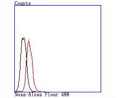
Fig3: Flow cytometric analysis of Jurkat cells with Noxa antibody at 1/100 dilution (red) compared with an unlabelled control (cells without incubation with primary antibody; black).Application
Fig3: Flow cytometric analysis of Jurkat cells with Noxa antibody at 1/100 dilution (red) compared with an unlabelled control (cells without incubation with primary antibody; black).Application
Fig3: Flow cytometric analysis of Jurkat cells with Noxa antibody at 1/100 dilution (red) compared with an unlabelled control (cells without incubation with primary antibody; black).| Product Name | Noxa |
|---|---|
| Antibody Type | Primary Antibodies |
| Antigen Alias | Adult T cell leukemia derived PMA responsive antibody APR antibody APR_HUMAN antibody ATL-derived antibody Immediate early response protein APR antibody Immediate-early-response protein APR antibody NOXA antibody Phorbol 12 myristate 13 acetate induced protein 1 antibody Phorbol-12-myristate-13-acetate-induced protein 1 antibody PMA induced protein 1 antibody PMA-induced protein 1 antibody PMA-responsive gene antibody Pmaip1 antibody Protein Noxa antibody |
| Product description | Members of the Bcl-2 family of proteins interact to regulate programmed cell death (apoptosis) under a broad range of physiological conditions. Bcl-2, Bcl-xL, and several related proteins inhibit apoptosis, whereas other members of the Bcl-2 family, such as Bax and Bak, enhance cell death. NOXA, a pro-apoptotic member of the Bcl-2 family, contains the Bcl-2 homology 3 (BH3) region, but does not contain other BH domains. Murine cells constitutively express NOXA mRNA in small amounts in various organs; X-ray irradiation increases NOXA mRNA and protein expression levels. In human cells, NOXA, alternatively designated PMA-induced protein 1 or APR, displays high expression in the adult T cell leukemia cell line IKD, where it may function as an immediate-early-response gene. The NOXA protein selectively localizes to mitochondria. |
| Immunogen | recombinant protein |
| Clonality | Monoclonal |
|---|---|
| Isotype | IgG |
| Host Species | Recombinant rabbit |
| Tested Applications | WBIHCFC |
| WB:1:500 IHC:1:50-1:200 FC:1:50 |
|
| Species Reactivity | Human |
| Concentration | 1mg/ml |
| Alternative Names | Adult T cell leukemia derived PMA responsive antibody APR antibody APR_HUMAN antibody ATL-derived antibody Immediate early response protein APR antibody Immediate-early-response protein APR antibody NOXA antibody Phorbol 12 myristate 13 acetate induced protein 1 antibody Phorbol-12-myristate-13-acetate-induced protein 1 antibody PMA induced protein 1 antibody PMA-induced protein 1 antibody PMA-responsive gene antibody Pmaip1 antibody Protein Noxa antibody |
|---|---|
| Molecular Weight(MW) | 6 kDa |
| Cellular Localization | Mitochondrion. |
| SwissProt ID | Q13794 |
|---|
Application
Fig3: Flow cytometric analysis of Jurkat cells with Noxa antibody at 1/100 dilution (red) compared with an unlabelled control (cells without incubation with primary antibody; black).
Application
Fig3: Flow cytometric analysis of Jurkat cells with Noxa antibody at 1/100 dilution (red) compared with an unlabelled control (cells without incubation with primary antibody; black).
Application
Fig3: Flow cytometric analysis of Jurkat cells with Noxa antibody at 1/100 dilution (red) compared with an unlabelled control (cells without incubation with primary antibody; black).| Positive Control | Human liver tissue, human spleen tissue, human kidney tissue, Jurkat. |
|---|---|
| Application Notes | WB:1:500 IHC:1:50-1:200 FC:1:50 |
| Form | Liquid |
|---|---|
| Storage Instructions | Store at +4℃ after thawing. Aliquot store at -20℃ or -80℃. Avoid repeated freeze / thaw cycles. |
| Storage Buffer | 1*TBS (pH7.4), 1%BSA, 40%Glycerol. Preservative: 0.05% Sodium Azide. |


