Application
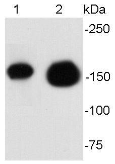
Fig1: Western blot analysis of ErbB2 on different cell lysates using anti- ErbB2 antibody at 1/500 dilution. Positive control: Lane 1: SKOV-3 Lane2: SKBR-3Application
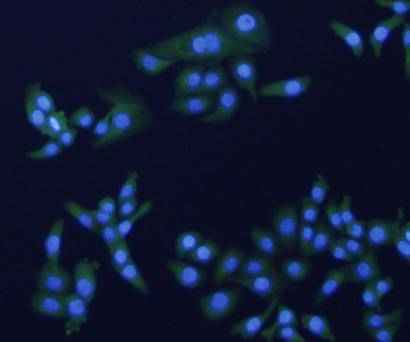
Fig2: ICC staining ErbB2 in SKOV-3 cells (green). The nuclear counter stain is DAPI (blue). Cells were fixed in paraformaldehyde, permeabilised with 0.25% Triton X100/PBS.Application
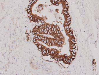
Fig3: Immunohistochemical analysis of paraffin-embedded human stomach carcinoma tissue using anti-Her 2 antibody. Counter stained with hematoxylin.| Product Name | HER2 / ErbB2 |
|---|---|
| Antibody Type | Primary Antibodies |
| Antigen Alias | Verb b2 erythroblastic leukemia viral oncogene homolog 2, neuro/glioblastoma derived oncogene homolog antibody C erb B2/neu protein antibody CD340 antibody CD340 antigen antibody Cerb B2/neu protein antibody CerbB2 antibody Erb b2 receptor tyrosine kinase 2 antibody ERBB2 antibody ERBB2_HUMAN antibody HER 2 antibody HER 2/NEU antibody HER2 antibody Herstatin antibody Human epidermal growth factor receptor 2 antibody Metastatic lymph node gene 19 protein antibody MLN 19 antibody MLN19 antibody NEU antibody NEU proto oncogene antibody Neuro/glioblastoma derived oncogene homolog antibody Neuroblastoma/glioblastoma derived oncogene homolog antibody NGL antibody p185erbB2 antibody Proto-oncogene c-ErbB-2 antibody Proto-oncogene Neu antibody Receptor tyrosine-protein kinase erbB-2 antibody TKR1 antibody Tyrosine kinase type cell surface receptor HER2 antibody Tyrosine kinase-type cell surface receptor HER2 antibody V erb b2 avian erythroblastic leukemia viral oncogene homolog 2 (neuro/glioblastoma derived oncogene homolog) antibody V erb b2 avian erythroblastic leukemia viral oncogene homolog 2 antibody V erb b2 avian erythroblastic leukemia viral oncoprotein 2 antibody V erb b2 erythroblastic leukemia viral oncogene homolog 2, neuro/glioblastoma derived oncogene homolog (avian) antibody V erb b2 erythroblastic leukemia viral oncogene homolog 2, neuro/glioblastoma derived oncogene homolog antibody Verb b2 erythroblastic leukemia viral oncogene homolog 2, neuro/glioblastoma derived oncogene homolog (avian) antibody |
| Product description | HER2 is a member of the human epidermal growth factor receptor (HER/EGFR/ERBB) family. Amplification or overexpression of this oncogene has been shown to play an important role in the development and progression of certain aggressive types of breast cancer. In recent years the protein has become an important biomarker and target of therapy for approx. 30% of breast cancer patients. Over-expression is also known to occur in ovarian, stomach, and aggressive forms of uterine cancer, such as uterine serous endometrial carcinoma. The expression of HER2 is regulated by signaling through estrogen receptors. Normally, estradiol and tamoxifen acting through the estrogen receptor down-regulate the expression of HER2. However, when the ratio of the coactivator AIB-3 exceeds that of the corepressor PAX2, the expression of HER2 is upregulated in the presence of tamoxifen, leading to tamoxifen-resistant breast cancer. |
| Immunogen | peptide |
| Clonality | Polyclonal |
|---|---|
| Isotype | IgG |
| Host Species | Rabbit |
| Tested Applications | WBICCIHC |
| WB:1:500-1:1,000 ICC:1:200 IHC:1:200 |
|
| Species Reactivity | Human |
| Concentration | 1mg/ml |
| Alternative Names | Verb b2 erythroblastic leukemia viral oncogene homolog 2 neuro/glioblastoma derived oncogene homolog antibody C erb B2/neu protein antibody CD340 antibody CD340 antigen antibody Cerb B2/neu protein antibody CerbB2 antibody Erb b2 receptor tyrosine kinase 2 antibody ERBB2 antibody ERBB2_HUMAN antibody HER 2 antibody HER 2/NEU antibody HER2 antibody Herstatin antibody Human epidermal growth factor receptor 2 antibody Metastatic lymph node gene 19 protein antibody MLN 19 antibody MLN19 antibody NEU antibody NEU proto oncogene antibody Neuro/glioblastoma derived oncogene homolog antibody Neuroblastoma/glioblastoma derived oncogene homolog antibody NGL antibody p185erbB2 antibody Proto-oncogene c-ErbB-2 antibody Proto-oncogene Neu antibody Receptor tyrosine-protein kinase erbB-2 antibody TKR1 antibody Tyrosine kinase type cell surface receptor HER2 antibody Tyrosine kinase-type cell surface receptor HER2 antibody V erb b2 avian erythroblastic leukemia viral oncogene homolog 2 (neuro/glioblastoma derived oncogene homolog) antibody V erb b2 avian erythroblastic leukemia viral oncogene homolog 2 antibody V erb b2 avian erythroblastic leukemia viral oncoprotein 2 antibody V erb b2 erythroblastic leukemia viral oncogene homolog 2 neuro/glioblastoma derived oncogene homolog (avian) antibody V erb b2 erythroblastic leukemia viral oncogene homolog 2 neuro/glioblastoma derived oncogene homolog antibody Verb b2 erythroblastic leukemia viral oncogene homolog 2 neuro/glioblastoma derived oncogene homolog (avian) antibody |
|---|---|
| Molecular Weight(MW) | 140kDa |
| Cellular Localization | Cell membrane |
| SwissProt ID | P04626 |
|---|
Application
Fig1: Western blot analysis of ErbB2 on different cell lysates using anti- ErbB2 antibody at 1/500 dilution. Positive control: Lane 1: SKOV-3 Lane2: SKBR-3
Application
Fig2: ICC staining ErbB2 in SKOV-3 cells (green). The nuclear counter stain is DAPI (blue). Cells were fixed in paraformaldehyde, permeabilised with 0.25% Triton X100/PBS.
Application
Fig3: Immunohistochemical analysis of paraffin-embedded human stomach carcinoma tissue using anti-Her 2 antibody. Counter stained with hematoxylin.| Positive Control | SKOV-3, SKBR-3, human stomach carcinoma tissue |
|---|---|
| Application Notes | WB:1:500-1:1,000 ICC:1:200 IHC:1:200 |
| Form | Liquid |
|---|---|
| Storage Instructions | Store at +4℃ after thawing. Aliquot store at -20℃ or -80℃. Avoid repeated freeze / thaw cycles. |
| Storage Buffer | 1*TBS (pH7.4), 0.5%BSA, 40%Glycerol. Preservative: 0.05% Sodium Azide. |


