Application
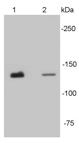
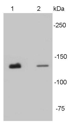
Fig1: Western blot analysis of N-cadherin on different tissue lysates using anti- N-cadherin antibody at 1/500 dilution. Positive control: Lane 1: Mouse heart Lane2 : Human heartApplication
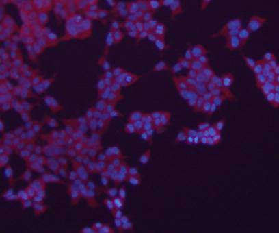
Fig2: ICC staining N-Cadherin in 293 cells (red). The nuclear counter stain is DAPI (blue). Cells were Cells were fixed in paraformaldehyde, permeabilised with 0.25% Triton X100/PBS.Application
Fig3: ICC staining N-Cadherin in mouse embryonic stem cells (green). The nuclear counter stain is DAPI (blue). Cells were Cells were fixed in paraformaldehyde, permeabilised with 0.25% Triton X100/PBS.Application
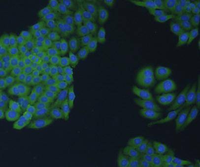
Fig4: ICC staining N-Cadherin in HepG2 cells (green). The nuclear counter stain is DAPI (blue). Cells were Cells were fixed in paraformaldehyde, permeabilised with 0.25% Triton X100/PBS.Application
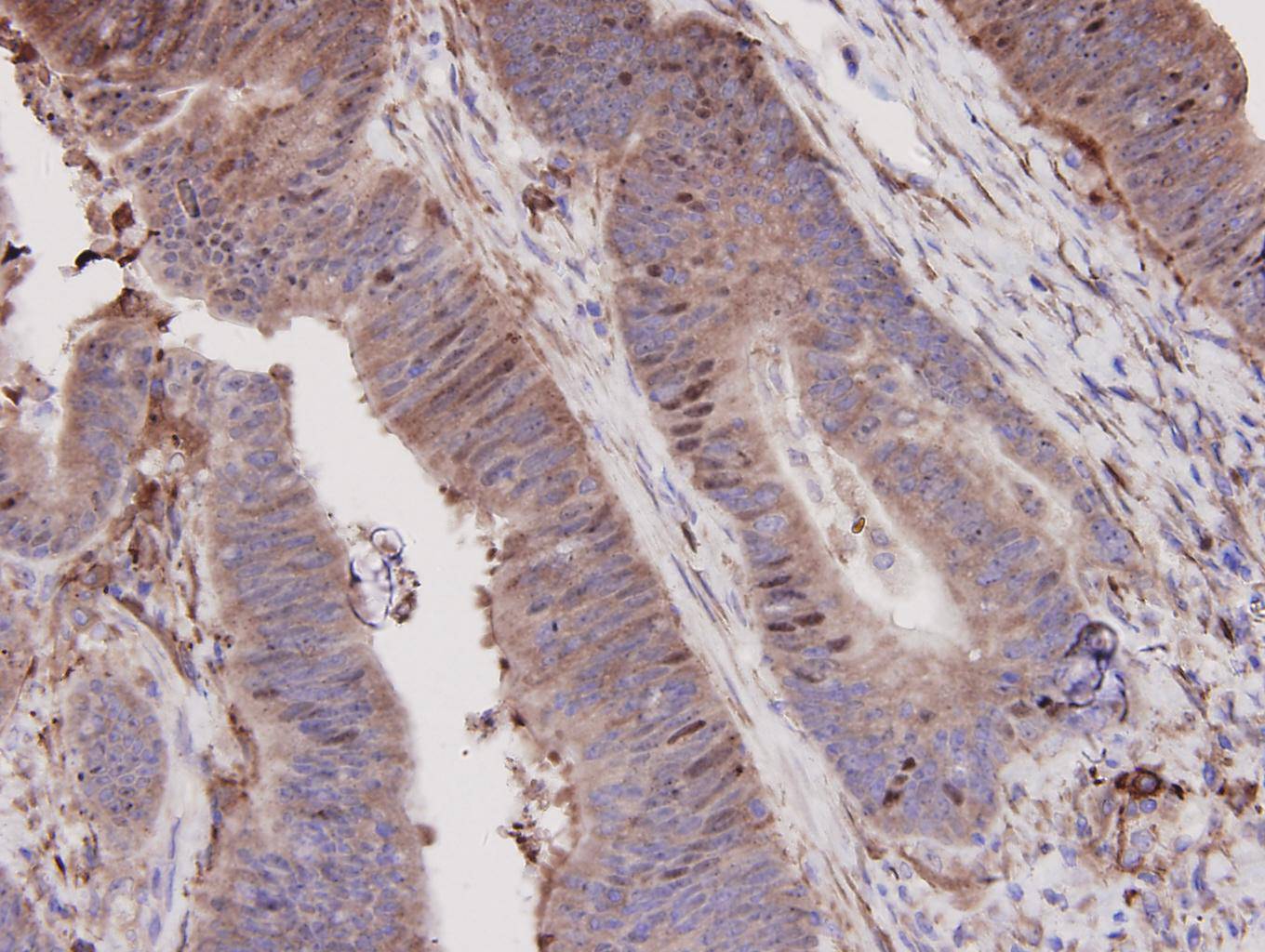
Fig5: Immunohistochemical analysis of paraffin-embedded human colon carcinoma tissue using anti-N-Cadherin antibody. Counter stained with hematoxylin.Application
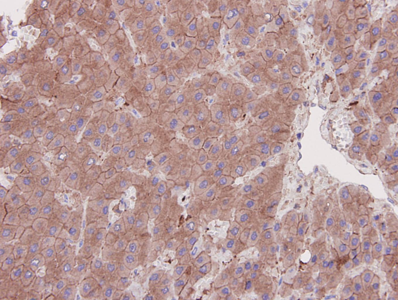
Fig6: Immunohistochemical analysis of paraffin-embedded human liver carcinoma tissue using anti-N-Cadherin antibody. Counter stained with hematoxylin.| Product Name | N-Cadherin |
|---|---|
| Antibody Type | Primary Antibodies |
| Antigen Alias | CADH2_HUMAN antibody Cadherin 2 antibody Cadherin 2 N cadherin neuronal antibody Cadherin 2 type 1 antibody Cadherin 2 type 1 N cadherin neuronal antibody Cadherin 2, type 1, N-cadherin (neuronal) antibody Cadherin-2 antibody Cadherin2 antibody Calcium dependent adhesion protein neuronal antibody CD325 antibody CD325 antigen antibody CDH2 antibody CDHN antibody CDw325 antibody CDw325 antigen antibody N cadherin 1 antibody N-cadherin antibody NCAD antibody Neural cadherin antibody OTTHUMP00000066304 antibody OTTHUMP00000067378 antibody |
| Product description | The cadherins are a large family of cell adhesion molecules involved in calcium-dependent recognition and adhesion events. The cadherin subfamily includes N-, P-, B-, and E- cadherins, and other members. Recent studies indicate that cancer cells have up-regulated N-cadherin in addition to loss of E-cadherin. N-cadherin cooperates with the FGF receptor, leading to overexpression of MMP-9 and cellular invasion. N-Cadherin is commonly found in cancer cells and provides a mechanism for transendothelial migration. When a cancer cell adheres to the endothelial cells of a blood vessel it up-regulates the src kinase pathway, which phosphorylates beta-catenins attached to both N-cadherin (this protein) and E-cadherins. |
| Immunogen | Synthetic peptide (KLH-coupled) within human N-cadherin 360-420 aa. |
| Clonality | Polyclonal |
|---|---|
| Isotype | IgG |
| Host Species | Rabbit |
| Tested Applications | WBICCIHCFC |
| WB:1:500 ICC:1:200 IHC:1:200 FC:1:50 |
|
| Species Reactivity | HumanMouse |
| Concentration | 1mg/ml |
| Alternative Names | CADH2_HUMAN antibody
Cadherin 2 antibody
Cadherin 2 N cadherin neuronal antibody
Cadherin 2 type 1 antibody
Cadherin 2 type 1 N cadherin neuronal antibody
Cadherin 2 type 1 N-cadherin (neuronal) antibody Cadherin-2 antibody Cadherin2 antibody Calcium dependent adhesion protein neuronal antibody CD325 antibody CD325 antigen antibody CDH2 antibody CDHN antibody CDw325 antibody CDw325 antigen antibody N cadherin 1 antibody N-cadherin antibody NCAD antibody Neural cadherin antibody OTTHUMP00000066304 antibody OTTHUMP00000067378 antibody |
|---|---|
| Molecular Weight(MW) | 140 kDa |
| Cellular Localization | Cell membrane |
Application
Fig1: Western blot analysis of N-cadherin on different tissue lysates using anti- N-cadherin antibody at 1/500 dilution. Positive control: Lane 1: Mouse heart Lane2 : Human heart
Application
Fig2: ICC staining N-Cadherin in 293 cells (red). The nuclear counter stain is DAPI (blue). Cells were Cells were fixed in paraformaldehyde, permeabilised with 0.25% Triton X100/PBS.
Application
Fig3: ICC staining N-Cadherin in mouse embryonic stem cells (green). The nuclear counter stain is DAPI (blue). Cells were Cells were fixed in paraformaldehyde, permeabilised with 0.25% Triton X100/PBS.
Application
Fig4: ICC staining N-Cadherin in HepG2 cells (green). The nuclear counter stain is DAPI (blue). Cells were Cells were fixed in paraformaldehyde, permeabilised with 0.25% Triton X100/PBS.
Application
Fig5: Immunohistochemical analysis of paraffin-embedded human colon carcinoma tissue using anti-N-Cadherin antibody. Counter stained with hematoxylin.
Application
Fig6: Immunohistochemical analysis of paraffin-embedded human liver carcinoma tissue using anti-N-Cadherin antibody. Counter stained with hematoxylin.| Positive Control | Mouse heart, human heart, 293, mouse embryonic stem cell, HepG2 Human colon?carcinoma tissue, human liver?carcinoma tissue |
|---|---|
| Application Notes | WB:1:500 ICC:1:200 IHC:1:200 FC:1:50 |
| Form | Liquid |
|---|---|
| Storage Instructions | Store at +4℃ after thawing. Aliquot store at -20℃. Avoid repeated freeze / thaw cycles. |
| Storage Buffer | 1*TBS (pH7.4), 1%BSA, 40%Glycerol. Preservative: 0.05% Sodium Azide. |


