Application
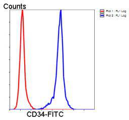
Fig5: Flow cytometric analysis of Jurkat cells with CD34 antibody at 1/50 dilution (blue) compared with an unlabelled control (cells without incubation with primary antibody; red). Goat anti rabbit IgG (FITC) was used as the secondary antibody.Application
Fig5: Flow cytometric analysis of Jurkat cells with CD34 antibody at 1/50 dilution (blue) compared with an unlabelled control (cells without incubation with primary antibody; red). Goat anti rabbit IgG (FITC) was used as the secondary antibody.Application
Fig5: Flow cytometric analysis of Jurkat cells with CD34 antibody at 1/50 dilution (blue) compared with an unlabelled control (cells without incubation with primary antibody; red). Goat anti rabbit IgG (FITC) was used as the secondary antibody.Application
Fig5: Flow cytometric analysis of Jurkat cells with CD34 antibody at 1/50 dilution (blue) compared with an unlabelled control (cells without incubation with primary antibody; red). Goat anti rabbit IgG (FITC) was used as the secondary antibody.Application
Fig5: Flow cytometric analysis of Jurkat cells with CD34 antibody at 1/50 dilution (blue) compared with an unlabelled control (cells without incubation with primary antibody; red). Goat anti rabbit IgG (FITC) was used as the secondary antibody.| Product Name | CD34 |
|---|---|
| Antibody Type | Primary Antibodies |
| Antigen Alias | CD34 antibody CD34 antigen antibody CD34 molecule antibody CD34_HUMAN antibody Cluster designation 34 antibody Hematopoietic progenitor cell antigen CD34 antibody HPCA1 antibody Mucosialin antibody OTTHUMP00000034733 antibody OTTHUMP00000034734 antibody |
| Product description | CD34 is a heavily glycosylated type I transmembrane molecule, that can be phoshorylated by a variety of kinases including Protein kinase C and Tyrosine kinases. CD34 antigen is expressed on small vessel endothelial cells and tumors of epithelial origin. CD34 is possibly an adhesion molecule with a putative role in early hematopoiesis by mediating the attachment of stem cells to the bone marrow extracellular matrix or directly to stromal cells. It could act as a scaffold for the attachment of lineage specific glycans, allowing stem cells to bind to lectins expressed by stromal cells or other marrow components. So CD34 is considered to be ideal marker for identifying and quantifying hematopoietic progenitor stem cells. With interaction with CrkL, CD34 becomes a substrate for PKC and CD34 surface expression is associated with activation of PKC. |
| Immunogen | peptide |
| Clonality | Polyclonal |
|---|---|
| Isotype | IgG |
| Host Species | Rabbit |
| Tested Applications | WBICCIHCFC |
| WB:1:1,000-1:2,000 ICC:1:200 IHC:1:2,000-1:3,000 FC:1:50-1:100 |
|
| Species Reactivity | HumanMouseRat |
| Concentration | 1mg/ml |
| Alternative Names | CD34 antibody
CD34 antigen antibody CD34 molecule antibody CD34_HUMAN antibody Cluster designation 34 antibody Hematopoietic progenitor cell antigen CD34 antibody HPCA1 antibody Mucosialin antibody OTTHUMP00000034733 antibody OTTHUMP00000034734 antibody |
|---|---|
| Molecular Weight(MW) | 120 kDa |
| Cellular Localization | Cell membrane |
| SwissProt ID | P28906 |
|---|
Application
Fig5: Flow cytometric analysis of Jurkat cells with CD34 antibody at 1/50 dilution (blue) compared with an unlabelled control (cells without incubation with primary antibody; red). Goat anti rabbit IgG (FITC) was used as the secondary antibody.
Application
Fig5: Flow cytometric analysis of Jurkat cells with CD34 antibody at 1/50 dilution (blue) compared with an unlabelled control (cells without incubation with primary antibody; red). Goat anti rabbit IgG (FITC) was used as the secondary antibody.
Application
Fig5: Flow cytometric analysis of Jurkat cells with CD34 antibody at 1/50 dilution (blue) compared with an unlabelled control (cells without incubation with primary antibody; red). Goat anti rabbit IgG (FITC) was used as the secondary antibody.
Application
Fig5: Flow cytometric analysis of Jurkat cells with CD34 antibody at 1/50 dilution (blue) compared with an unlabelled control (cells without incubation with primary antibody; red). Goat anti rabbit IgG (FITC) was used as the secondary antibody.
Application
Fig5: Flow cytometric analysis of Jurkat cells with CD34 antibody at 1/50 dilution (blue) compared with an unlabelled control (cells without incubation with primary antibody; red). Goat anti rabbit IgG (FITC) was used as the secondary antibody.| Positive Control | Human brain, mouse brain, mouse testis, moue thymus, mouse spleen, Jurkat, TF-1, F9, human embryonic stem cell, human tonsil tissue, human liver carcinoma tissue |
|---|---|
| Application Notes | WB:1:1,000-1:2,000 ICC:1:200 IHC:1:2,000-1:3,000 FC:1:50-1:100 |
| Form | Liquid |
|---|---|
| Storage Instructions | Store at +4℃ after thawing. Aliquot store at -20℃ or -80℃. Avoid repeated freeze / thaw cycles. |
| Storage Buffer | 1*TBS (pH7.4), 1%BSA, 40%Glycerol. Preservative: 0.05% Sodium Azide. |


