Application
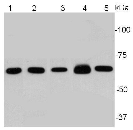
Fig1: Western blot analysis of NF-κB p65 on different lysates using anti-NF-κB p65 antibody at 1/1000 dilution.Positive control:Lane1: HelaLane2:A549Lane3: PC12Lane 4: Mouse embryonic stem cellLane5: NIH/3T3Application
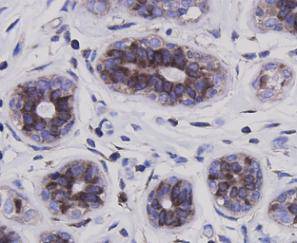
Fig2: Immunohistochemical analysis of paraffin-embedded human breast carcinoma tissue using anti-NF-κB p65 antibody. Counter stained with hematoxylin.Application
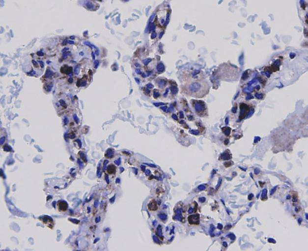
Fig3: Immunohistochemical analysis of paraffin-embedded human lung carcinoma tissue using anti-NF-κB p65 antibody. Counter stained with hematoxylin.Application
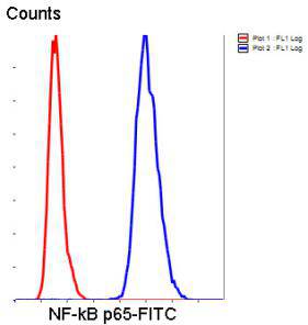
Fig4: Flow cytometric analysis of Hela cells with NF-κB p65 antibody at 1/50 dilution (blue) compared with an unlabelled control (cells without incubation with primary antibody; red). Goat anti rabbit IgG (FITC) was used as the secondary antibody.| Product Name | NF-κB p65 |
|---|---|
| Antibody Type | Primary Antibodies |
| Antigen Alias | Avian reticuloendotheliosis viral (v rel) oncogene homolog A antibody MGC131774 antibody NF kappa B p65delta3 antibody NFKB3 antibody Nuclear Factor NF Kappa B p65 Subunit antibody Nuclear factor NF-kappa-B p65 subunit antibody Nuclear factor of kappa light polypeptide gene enhancer in B cells 3 antibody Nuclear factor of kappa light polypeptide gene enhancer in B-cells 3 antibody OTTHUMP00000233473 antibody OTTHUMP00000233474 antibody OTTHUMP00000233475 antibody OTTHUMP00000233476 antibody OTTHUMP00000233900 antibody p65 antibody p65 NF kappaB antibody p65 NFkB antibody relA antibody TF65_HUMAN antibody Transcription factor p65 antibody v rel avian reticuloendotheliosis viral oncogene homolog A (nuclear factor of kappa light polypeptide gene enhancer in B cells 3 (p65)) antibody V rel avian reticuloendotheliosis viral oncogene homolog A antibody v rel reticuloendotheliosis viral oncogene homolog A (avian) antibody V rel reticuloendotheliosis viral oncogene homolog A, nuclear factor of kappa light polypeptide gene enhancer in B cells 3, p65 antibody |
| Product description | NF-kappa-B is a pleiotropic transcription factor present in almost all cell types and is the endpoint of a series of signal transduction events that are initiated by a vast array of stimuli related to many biological processes such as inflammation, immunity, differentiation, cell growth, tumorigenesis and apoptosis. NF-kappa-B is a homo- or heterodimeric complex formed by the Rel-like domain-containing proteins RELA/p65, RELB, NFKB1/p105, NFKB1/p50, REL and NFKB2/p52 and the heterodimeric p65-p50 complex appears to be most abundant one. In unstimulated cells, NF-κB is sequestered in the cytoplasm by IκB inhibitory proteins. NF-κB-activating agents can induce the phosphorylation of IκB proteins, targeting them for rapid degradation through the ubiquitin-proteasome pathway and releasing NF-κB to enter the nucleus where it regulates gene expression. |
| Immunogen | This antibody is produced by immunizing rabbits with a synthetic peptide (KLH-coupled) corresponding to N-terminal NF-κB p65. |
| Clonality | Polyclonal |
|---|---|
| Isotype | IgG |
| Host Species | Rabbit |
| Tested Applications | WBIHCFC |
| WB:1:1,000-1:2,000 IHC:1:100-1:200 FC:1:50-1:100 |
|
| Species Reactivity | HumanMouseRat |
| Concentration | 1mg/ml |
| Alternative Names | Avian reticuloendotheliosis viral (v rel) oncogene homolog A antibody
MGC131774 antibody
NF kappa B p65delta3 antibody
NFKB3 antibody
Nuclear Factor NF Kappa B p65 Subunit antibody
Nuclear factor NF-kappa-B p65 subunit antibody
Nuclear factor of kappa light polypeptide gene enhancer in B cells 3 antibody
Nuclear factor of kappa light polypeptide gene enhancer in B-cells 3 antibody
OTTHUMP00000233473 antibody
OTTHUMP00000233474 antibody
OTTHUMP00000233475 antibody
OTTHUMP00000233476 antibody
OTTHUMP00000233900 antibody
p65 antibody
p65 NF kappaB antibody
p65 NFkB antibody
relA antibody
TF65_HUMAN antibody
Transcription factor p65 antibody
v rel avian reticuloendotheliosis viral oncogene homolog A (nuclear factor of kappa light polypeptide gene enhancer in B cells 3 (p65)) antibody
V rel avian reticuloendotheliosis viral oncogene homolog A antibody
v rel reticuloendotheliosis viral oncogene homolog A (avian) antibody
V rel reticuloendotheliosis viral oncogene homolog A nuclear factor of kappa light polypeptide gene enhancer in B cells 3 p65 antibody |
|---|---|
| Molecular Weight(MW) | 65 kDa |
| Cellular Localization | Nucleus, cytoplasm |
Application
Fig1: Western blot analysis of NF-κB p65 on different lysates using anti-NF-κB p65 antibody at 1/1000 dilution.Positive control:Lane1: HelaLane2:A549Lane3: PC12Lane 4: Mouse embryonic stem cellLane5: NIH/3T3
Application
Fig2: Immunohistochemical analysis of paraffin-embedded human breast carcinoma tissue using anti-NF-κB p65 antibody. Counter stained with hematoxylin.
Application
Fig3: Immunohistochemical analysis of paraffin-embedded human lung carcinoma tissue using anti-NF-κB p65 antibody. Counter stained with hematoxylin.
Application
Fig4: Flow cytometric analysis of Hela cells with NF-κB p65 antibody at 1/50 dilution (blue) compared with an unlabelled control (cells without incubation with primary antibody; red). Goat anti rabbit IgG (FITC) was used as the secondary antibody.| Positive Control | Hela, A549, PC12, mouse embryonic stem cell NIH/3T3, human breast carcinoma tissue, human lung carcinoma tissue |
|---|---|
| Application Notes | WB:1:1,000-1:2,000 IHC:1:100-1:200 FC:1:50-1:100 |
| Form | Liquid |
|---|---|
| Storage Instructions | Store at +4℃ after thawing. Aliquot store at -20℃. Avoid repeated freeze / thaw cycles. |
| Storage Buffer | 1*TBS (pH7.4), 1%BSA, 40%Glycerol. Preservative: 0.05% Sodium Azide. |


