Application
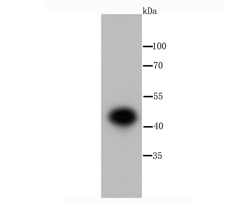
Fig1: Western blot analysis of IKK gamma on Hela cell using anti-IKK gamma antibody at 1/1,000 dilution.Application
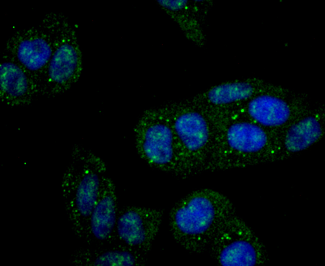
Fig2: ICC staining IKK gamma in Hela cells (green). The nuclear counter stain is DAPI (blue). Cells were fixed in paraformaldehyde, permeabilised with 0.25% Triton X100/PBS.Application
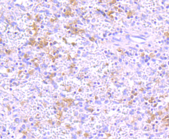
Fig3: Immunohistochemical analysis of paraffin-embedded human spleen tissue using anti-IKK gamma antibody. Counter stained with hematoxylin.Application
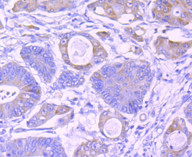
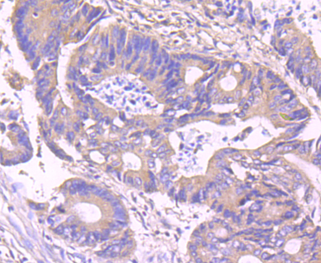
Fig4: Immunohistochemical analysis of paraffin-embedded human colon cancer tissue using anti-IKK gamma antibody. Counter stained with hematoxylin.Application
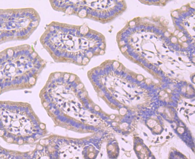
Fig5: Immunohistochemical analysis of paraffin-embedded mouse colon tissue using anti-IKK gamma antibody. Counter stained with hematoxylin.Application
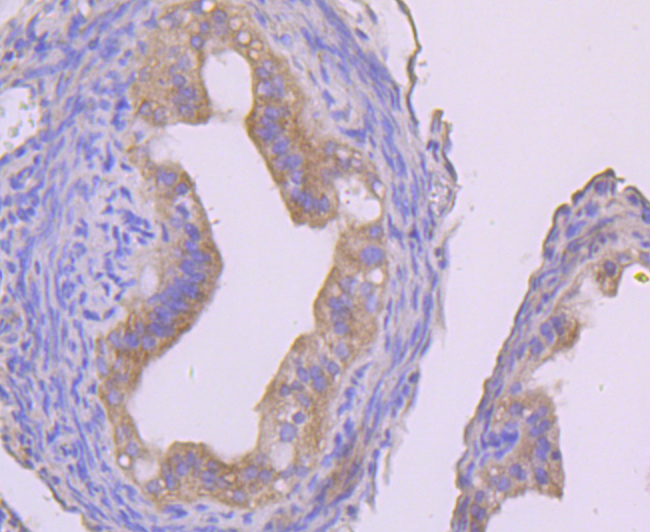
Fig6: Immunohistochemical analysis of paraffin-embedded mouse fallopian tube tissue using anti-IKK gamma antibody. Counter stained with hematoxylin.Application
Fig7: Immunohistochemical analysis of paraffin-embedded human colon tissue using anti-IKK gamma antibody. Counter stained with hematoxylin.Application
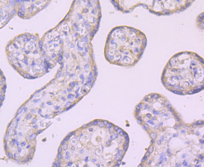
Fig8: Immunohistochemical analysis of paraffin-embedded human placenta tissue using anti-IKK gamma antibody. Counter stained with hematoxylin.Application
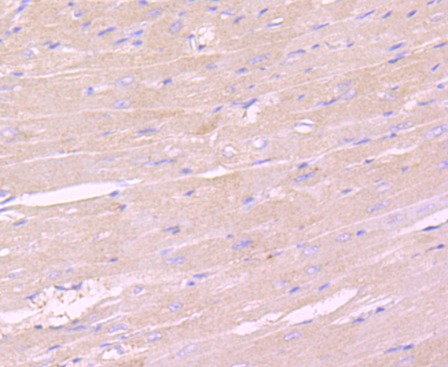
Fig9: Immunohistochemical analysis of paraffin-embedded mouse heart tissue using anti-IKK gamma antibody. Counter stained with hematoxylin.Application
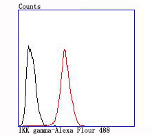
Fig10: Flow cytometric analysis of Hela cells with IKK gamma antibody at 1/100 dilution (red) compared with an unlabelled control (cells without incubation with primary antibody; black).| Product Name | IKK gamma [JA11-83] |
|---|---|
| Antibody Type | Primary Antibodies |
| Antigen Alias | IkB kinase associated protein 1 antibody IkB kinase subunit gamma antibody Inhibitor of nuclear factor kappa B kinase subunit gamma antibody AMCBX1 antibody FIP 3 antibody FIP-3 antibody FIP3 antibody Fip3p antibody I kappa B kinase gamma antibody I-kappa-B kinase subunit gamma antibody IkB kinase gamma subunit antibody IkB kinase subunit gamma antibody IkB kinase-associated protein 1 antibody Ikbkg antibody IKK-gamma antibody IKKAP1 antibody IKKG antibody IMD33 antibody Incontinentia pigmenti antibody Inhibitor of kappa light polypeptide gene enhancer in B cells, kinase gamma antibody Inhibitor of kappa light polypeptide gene enhancer in B cells, kinase of, gamma antibody Inhibitor of nuclear factor kappa-B kinase subunit gamma antibody IP antibody IP1 antibody IP2 antibody IPD2 antibody NEMO antibody NEMO_HUMAN antibody NF kappa B essential modifier antibody NF kappa B essential modulator antibody NF-kappa-B essential modifier antibody NF-kappa-B essential modulator antibody ZC2HC9 antibody |
| Product description | Regulatory subunit of the IKK core complex which phosphorylates inhibitors of NF-kappa-B thus leading to the dissociation of the inhibitor/NF-kappa-B complex and ultimately the degradation of the inhibitor. Its binding to scaffolding polyubiquitin seems to play a role in IKK activation by multiple signaling receptor pathways. However, the specific type of polyubiquitin recognized upon cell stimulation (either 'Lys-63'-linked or linear polyubiquitin) and its functional importance is reported conflictingly. Also considered to be a mediator for TAX activation of NF-kappa-B. Could be implicated in NF-kappa-B-mediated protection from cytokine toxicity. Essential for viral activation of IRF3. Involved in TLR3- and IFIH1-mediated antiviral innate response; this function requires 'Lys-27'-linked polyubiquitination. |
| Immunogen | recombinant protein |
| Clonality | Monoclonal |
|---|---|
| Isotype | IgG |
| Host Species | Recombinant rabbit |
| Tested Applications | WBIHCICC/IFIPFC |
| WB:1:500-1:2,000 ICC:1:50-1:200 IHC:1:20-1:50 FC:1:50-1:100 |
|
| Species Reactivity | HumanMouseRat |
| Concentration | 1mg/ml |
| Alternative Names | IkB kinase associated protein 1 antibody
IkB kinase subunit gamma antibody
Inhibitor of nuclear factor kappa B kinase subunit gamma antibody
AMCBX1 antibody
FIP 3 antibody
FIP-3 antibody
FIP3 antibody
Fip3p antibody
I kappa B kinase gamma antibody
I-kappa-B kinase subunit gamma antibody
IkB kinase gamma subunit antibody
IkB kinase subunit gamma antibody
IkB kinase-associated protein 1 antibody
Ikbkg antibody
IKK-gamma antibody
IKKAP1 antibody
IKKG antibody
IMD33 antibody
Incontinentia pigmenti antibody
Inhibitor of kappa light polypeptide gene enhancer in B cells kinase gamma antibody Inhibitor of kappa light polypeptide gene enhancer in B cells kinase of gamma antibody Inhibitor of nuclear factor kappa-B kinase subunit gamma antibody IP antibody IP1 antibody IP2 antibody IPD2 antibody NEMO antibody NEMO_HUMAN antibody NF kappa B essential modifier antibody NF kappa B essential modulator antibody NF-kappa-B essential modifier antibody NF-kappa-B essential modulator antibody ZC2HC9 antibody |
|---|---|
| Molecular Weight(MW) | 48 kDa |
| Cellular Localization | Cytoplasm. Nucleus. |
Application
Fig1: Western blot analysis of IKK gamma on Hela cell using anti-IKK gamma antibody at 1/1,000 dilution.
Application
Fig2: ICC staining IKK gamma in Hela cells (green). The nuclear counter stain is DAPI (blue). Cells were fixed in paraformaldehyde, permeabilised with 0.25% Triton X100/PBS.
Application
Fig3: Immunohistochemical analysis of paraffin-embedded human spleen tissue using anti-IKK gamma antibody. Counter stained with hematoxylin.
Application
Fig4: Immunohistochemical analysis of paraffin-embedded human colon cancer tissue using anti-IKK gamma antibody. Counter stained with hematoxylin.
Application
Fig5: Immunohistochemical analysis of paraffin-embedded mouse colon tissue using anti-IKK gamma antibody. Counter stained with hematoxylin.
Application
Fig6: Immunohistochemical analysis of paraffin-embedded mouse fallopian tube tissue using anti-IKK gamma antibody. Counter stained with hematoxylin.
Application
Fig7: Immunohistochemical analysis of paraffin-embedded human colon tissue using anti-IKK gamma antibody. Counter stained with hematoxylin.
Application
Fig8: Immunohistochemical analysis of paraffin-embedded human placenta tissue using anti-IKK gamma antibody. Counter stained with hematoxylin.
Application
Fig9: Immunohistochemical analysis of paraffin-embedded mouse heart tissue using anti-IKK gamma antibody. Counter stained with hematoxylin.
Application
Fig10: Flow cytometric analysis of Hela cells with IKK gamma antibody at 1/100 dilution (red) compared with an unlabelled control (cells without incubation with primary antibody; black).| Positive Control | Hela, human colon tissue, human spleen tissue, human colon cancer tissue,human placenta tissue, mouse colon tissue, mouse fallopian tube tissue, mouse heart tissue. |
|---|---|
| Application Notes | WB:1:500-1:2,000 ICC:1:50-1:200 IHC:1:20-1:50 FC:1:50-1:100 |
| Form | Liquid |
|---|---|
| Storage Instructions | Store at +4℃ after thawing. Aliquot store at -20℃ or -80℃. Avoid repeated freeze / thaw cycles. |
| Storage Buffer | 1*TBS (pH7.4), 1%BSA, 40%Glycerol. Preservative: 0.05% Sodium Azide. |


