Application
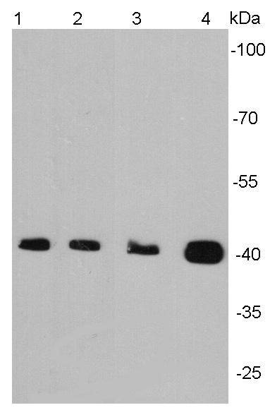
Fig1: Western blot analysis of alpha smooth muscle actin on different cell lysates using anti-Alpha smooth muscle actin antibody at 1/2000 dilution. Positive control: Lane 1: Jurkat Lane2 : NIH/3T3 Lane3: Mouse embryonic stem cell Lane 4: Mouse colonApplication
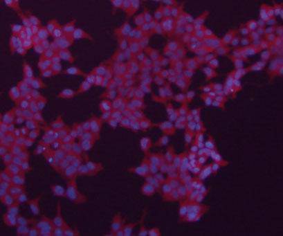
Fig2: ICC staining of Alpha smooth muscle actin in 293 cells (red). The nuclear counter stain is DAPI (blue). Cells were fixed in paraformaldehyde, permeabilised with 0.25% Triton X100/PBS.Application
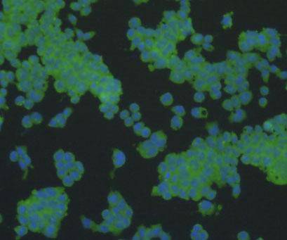
Fig3: ICC staining of Alpha smooth muscle actin in F9 cells (green). The nuclear counter stain is DAPI (blue). Cells were fixed in paraformaldehyde, permeabilised with 0.25% Triton X100/PBS.Application
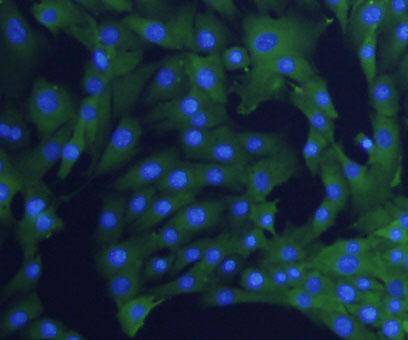
Fig4: ICC staining Alpha smooth muscle actin in NIH/3T3 cells (green). The nuclear counter stain is DAPI (blue). Cells were fixed in paraformaldehyde, permeabilised with 0.25% Triton X100/PBS.Application
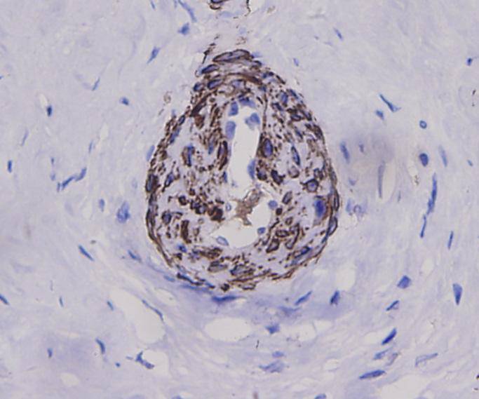
Fig5: Immunohistochemical analysis of paraffin-embedded human cervical tissue using anti-Alpha smooth muscle actin antibody. Counter stained with hematoxylin.Application
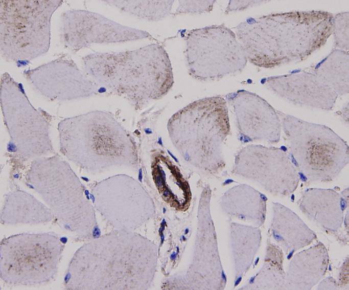
Fig6: Immunohistochemical analysis of paraffin-embedded mouse skeletal muscle tissue using anti-Alpha smooth muscle actin antibody. Counter stained with hematoxylin.Application
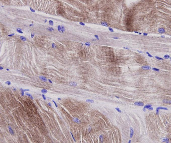
Fig7: Immunohistochemical analysis of paraffin-embedded mouse smooth muscle tissue using anti-Alpha smooth muscle actin antibody. Counter stained with hematoxylin.| Product Name | Alpha smooth muscle Actin |
|---|---|
| Antibody Type | Primary Antibodies |
| Antigen Alias | a actin antibody AAT6 antibody ACTA_HUMAN antibody ACTA2 antibody Actin alpha 2 smooth muscle aorta antibody Actin aortic smooth muscle antibody Actin, aortic smooth muscle antibody ACTSA antibody ACTVS antibody Alpha 2 actin antibody Alpha actin 2 antibody Alpha cardiac actin antibody Alpha-actin-2 antibody Cell growth inhibiting gene 46 protein antibody Cell growth-inhibiting gene 46 protein antibody GIG46 antibody Growth inhibiting gene 46 antibody MYMY5 antibody |
| Product description | Actins are highly conserved proteins involved in cell motility, structure and integrity. Actin has been found to be expressed in at least six isomeric forms. It is expressed in heart and skeletal striated muscle tissue, and in certain smooth muscle tissues, regulating contractile potentials for these cells. It is also expressed in the cytoplasm of non-muscle cells, functioning to control cell structure and motility. Smooth muscle alpha actin is of further interest because it is one of a few genes whose expression is relatively restricted to vascular smooth muscle cells. Expression of smooth muscle alpha actin is regulated by hormones, cell proliferation, and altered by pathological conditions including oncogenic transformation and atherosclerosis. |
| Immunogen | peptide |
| Clonality | Polyclonal |
|---|---|
| Isotype | IgG |
| Host Species | Rabbit |
| Tested Applications | WBICCIHC |
| WB:1:2,000 ICC:1:200 IHC:1:200 |
|
| Species Reactivity | HumanMouseRat |
| Concentration | 1mg/ml |
| Alternative Names | a actin antibody
AAT6 antibody
ACTA_HUMAN antibody
ACTA2 antibody
Actin alpha 2 smooth muscle aorta antibody
Actin aortic smooth muscle antibody
Actin aortic smooth muscle antibody ACTSA antibody ACTVS antibody Alpha 2 actin antibody Alpha actin 2 antibody Alpha cardiac actin antibody Alpha-actin-2 antibody Cell growth inhibiting gene 46 protein antibody Cell growth-inhibiting gene 46 protein antibody GIG46 antibody Growth inhibiting gene 46 antibody MYMY5 antibody |
|---|---|
| Molecular Weight(MW) | 42 kDa |
| Cellular Localization | Cytoskeleton |
Application
Fig1: Western blot analysis of alpha smooth muscle actin on different cell lysates using anti-Alpha smooth muscle actin antibody at 1/2000 dilution. Positive control: Lane 1: Jurkat Lane2 : NIH/3T3 Lane3: Mouse embryonic stem cell Lane 4: Mouse colon
Application
Fig2: ICC staining of Alpha smooth muscle actin in 293 cells (red). The nuclear counter stain is DAPI (blue). Cells were fixed in paraformaldehyde, permeabilised with 0.25% Triton X100/PBS.
Application
Fig3: ICC staining of Alpha smooth muscle actin in F9 cells (green). The nuclear counter stain is DAPI (blue). Cells were fixed in paraformaldehyde, permeabilised with 0.25% Triton X100/PBS.
Application
Fig4: ICC staining Alpha smooth muscle actin in NIH/3T3 cells (green). The nuclear counter stain is DAPI (blue). Cells were fixed in paraformaldehyde, permeabilised with 0.25% Triton X100/PBS.
Application
Fig5: Immunohistochemical analysis of paraffin-embedded human cervical tissue using anti-Alpha smooth muscle actin antibody. Counter stained with hematoxylin.
Application
Fig6: Immunohistochemical analysis of paraffin-embedded mouse skeletal muscle tissue using anti-Alpha smooth muscle actin antibody. Counter stained with hematoxylin.
Application
Fig7: Immunohistochemical analysis of paraffin-embedded mouse smooth muscle tissue using anti-Alpha smooth muscle actin antibody. Counter stained with hematoxylin.| Positive Control | Jurkat, NIH/3T3, mouse embryonic stem cell, mouse colon tissue, 293, F9, human cervical tissue, mouse skeletal muscle tissue, mouse smooth muscle tissue |
|---|---|
| Application Notes | WB:1:2,000 ICC:1:200 IHC:1:200 |
| Form | Liquid |
|---|---|
| Storage Instructions | Store at +4℃ after thawing. Aliquot store at -20℃ or -80℃. Avoid repeated freeze / thaw cycles. |
| Storage Buffer | 1*TBS (pH7.4), 1%BSA, 40%Glycerol. Preservative: 0.05% Sodium Azide. |


