Application
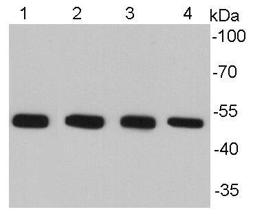
Fig1: Western blot analysis of Alpha-tubulin on different cell lysates using anti-Alpha -tubulin antibody at 1/10000 dilution. Positive control: Lane 1: NIH/3T3 Lane2: HepG2 Lane3: PC12 Lane 4: HelaApplication
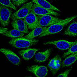
Fig2: ICC staining of Alpha-tubulin in HepG2 cells (green). The nuclear counter stain is DAPI (blue). Cells were fixed in paraformaldehyde, permeabilised with 0.25% Triton X100/PBS.Application
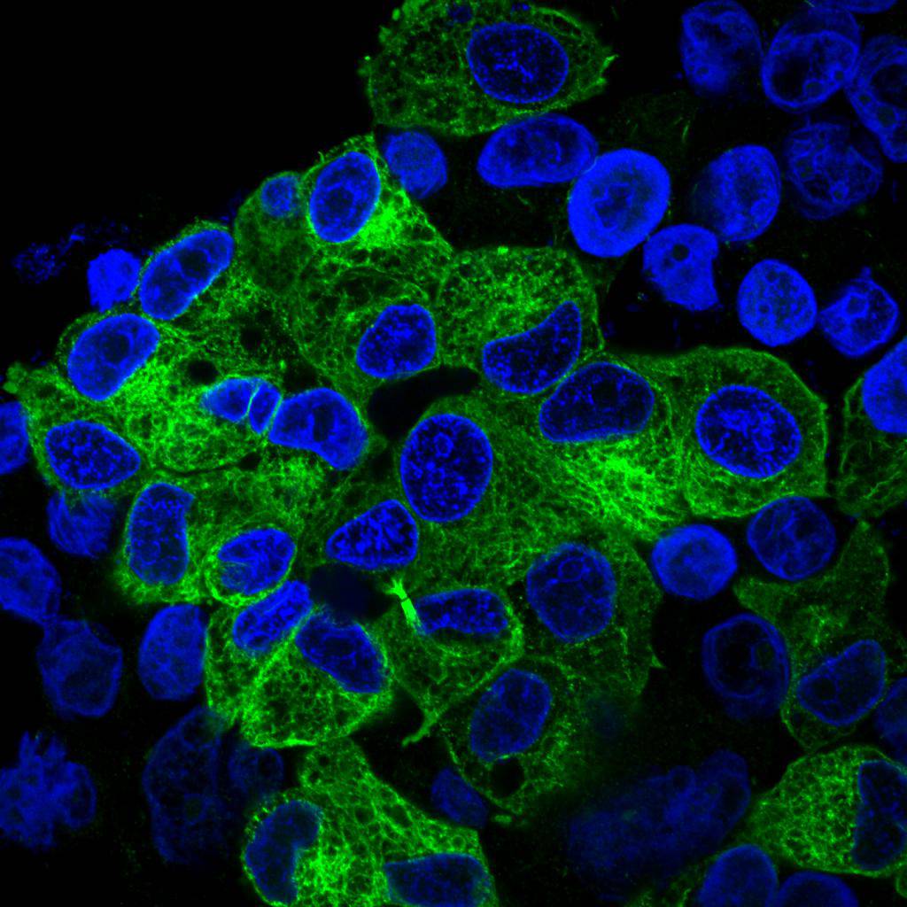
Fig3: ICC staining of Alpha-tubulin in Hela cells (green). The nuclear counter stain is DAPI (blue). Cells were fixed in paraformaldehyde, permeabilised with 0.25% Triton X100/PBS.Application
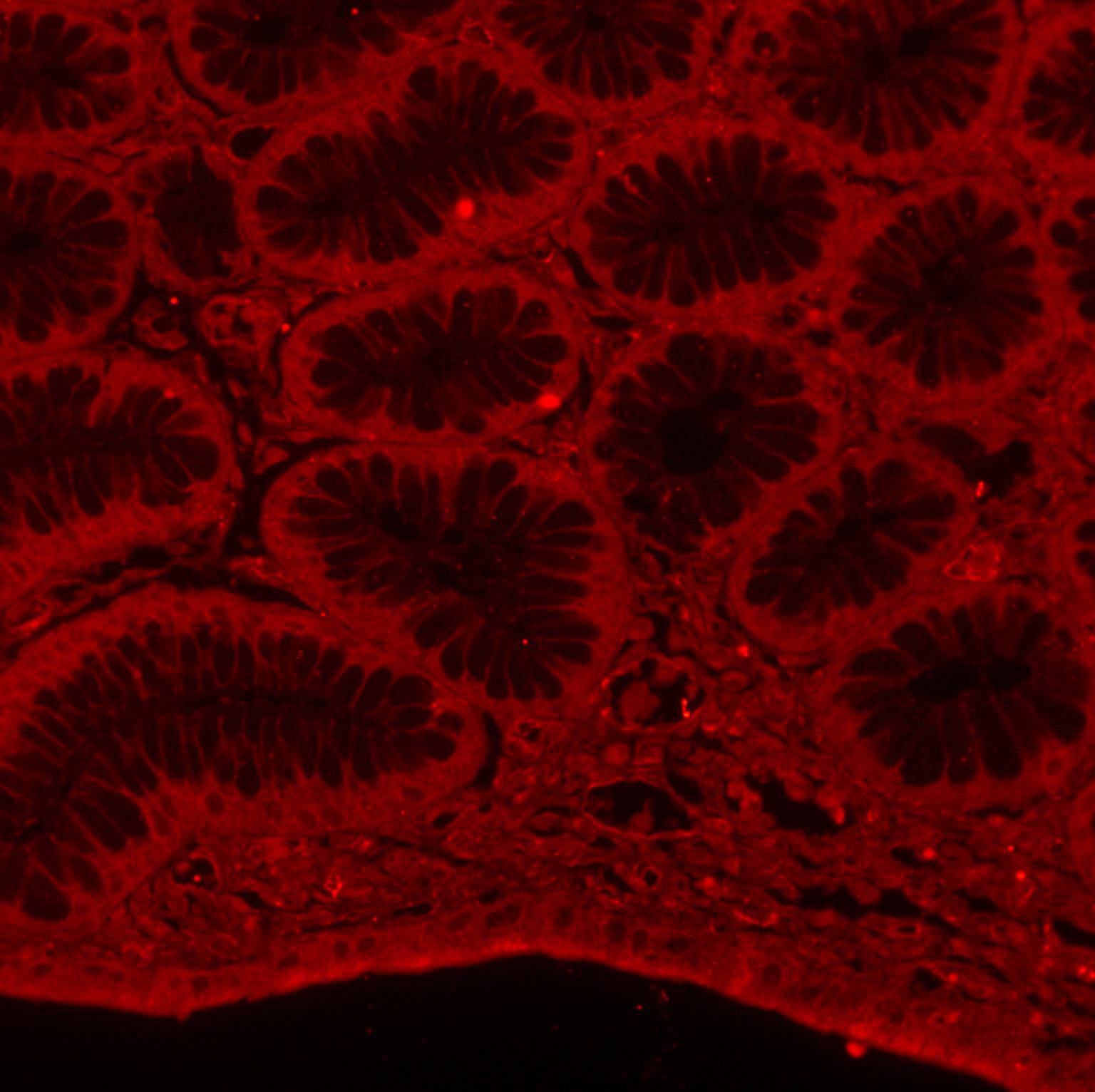
Fig4: IHC-P image of Alpha-tubulin staining with Alpha-tubulin antibody on tissue sections from human stomach. The secondary used was an Alexa-Fluor 555 conjugated goat anti-rabbit polyclonal.Application
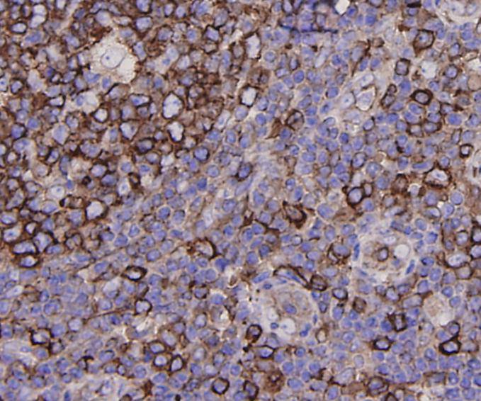
Fig5: Immunohistochemical analysis of paraffin-embedded human tonsil tissue using anti-Alpha-tubulin antibody. Counter stained with hematoxylin.Application
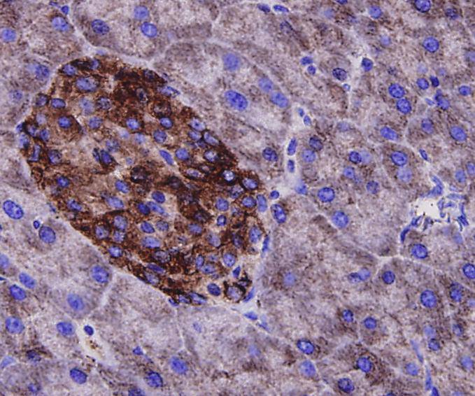
Fig6: Immunohistochemical analysis of paraffin-embedded mouse pancreas tissue using anti-Alpha-tubulin antibody. Counter stained with hematoxylin.Application
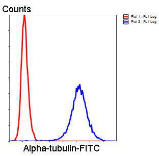
Fig7: Flow cytometric analysis of Hela cells with Alpha-tubulin antibody at 1/100 dilution (blue) compared with an unlabelled control (cells without incubation with primary antibody; red). Goat anti rabbit IgG (FITC) was used as the secondary antibody.| Product Name | Alpha-tubulin |
|---|---|
| Antibody Type | Primary Antibodies |
| Antigen Alias | TUBA1 antibody TUBA1A antibody TUBA2 antibody TUBA3 antibody TUBA3C antibody TUBA4A antibody Tubulin, alpha 1a antibody Tubulin, alpha 3c antibody Tubulin, alpha 4a antibody |
| Product description | The cytoskeleton consists of three types of cytosolic fibers: microtubules, microfilaments (actin filaments), and intermediate filaments. Globular tubulin subunits comprise the microtubule building block, with α/β-tubulin heterodimers forming the tubulin subunit common to all eukaryotic cells. Acetylation of alpha chains at Lys-40 stabilizes microtubules and affects affinity and processivity of microtubule motors. This modification has a role in multiple cellular functions, ranging from cell motility, cell cycle progression or cell differentiation to intracellular trafficking and signaling. |
| Immunogen | peptide |
| Clonality | Polyclonal |
|---|---|
| Isotype | IgG |
| Host Species | Rabbit |
| Tested Applications | WBIHCICCFCIF |
| WB:1:5,000-1:10,000 ICC:1:200-1:500 IHC:1:200-1:500 IF:1:200-1:500 FC:1:50-1:100 |
|
| Species Reactivity | HumanMouseRatZebra Fish |
| Concentration | 1mg/ml |
| Alternative Names | TUBA1 antibody
TUBA1A antibody
TUBA2 antibody
TUBA3 antibody
TUBA3C antibody
TUBA4A antibody
Tubulin alpha 1a antibody Tubulin alpha 3c antibody Tubulin alpha 4a antibody |
|---|---|
| Molecular Weight(MW) | 50 kDa |
| Cellular Localization | Cytoplasm ,cytoskeleton |
| SwissProt ID | P68366 |
|---|
Application
Fig1: Western blot analysis of Alpha-tubulin on different cell lysates using anti-Alpha -tubulin antibody at 1/10000 dilution. Positive control: Lane 1: NIH/3T3 Lane2: HepG2 Lane3: PC12 Lane 4: Hela
Application
Fig2: ICC staining of Alpha-tubulin in HepG2 cells (green). The nuclear counter stain is DAPI (blue). Cells were fixed in paraformaldehyde, permeabilised with 0.25% Triton X100/PBS.
Application
Fig3: ICC staining of Alpha-tubulin in Hela cells (green). The nuclear counter stain is DAPI (blue). Cells were fixed in paraformaldehyde, permeabilised with 0.25% Triton X100/PBS.
Application
Fig4: IHC-P image of Alpha-tubulin staining with Alpha-tubulin antibody on tissue sections from human stomach. The secondary used was an Alexa-Fluor 555 conjugated goat anti-rabbit polyclonal.
Application
Fig5: Immunohistochemical analysis of paraffin-embedded human tonsil tissue using anti-Alpha-tubulin antibody. Counter stained with hematoxylin.
Application
Fig6: Immunohistochemical analysis of paraffin-embedded mouse pancreas tissue using anti-Alpha-tubulin antibody. Counter stained with hematoxylin.
Application
Fig7: Flow cytometric analysis of Hela cells with Alpha-tubulin antibody at 1/100 dilution (blue) compared with an unlabelled control (cells without incubation with primary antibody; red). Goat anti rabbit IgG (FITC) was used as the secondary antibody.| Positive Control | NIH/3T3, Hela, HepG2, PC12, human stomach tissue, human tonsil tissue, mouse pancreas tissue |
|---|---|
| Application Notes | WB:1:5,000-1:10,000 ICC:1:200-1:500 IHC:1:200-1:500 IF:1:200-1:500 FC:1:50-1:100 |
| Form | Liquid |
|---|---|
| Storage Instructions | Store at +4℃ after thawing. Aliquot store at -20℃ or -80℃. Avoid repeated freeze / thaw cycles. |
| Storage Buffer | 1*TBS (pH7.4), 1%BSA, 40%Glycerol. Preservative: 0.05% Sodium Azide. |


