Application
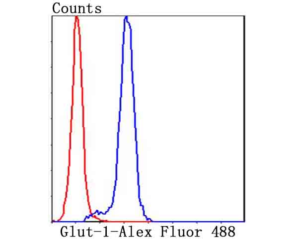
Fig6: Flow cytometric analysis of Hela cells with Glut1 antibody at 1/100 dilution (blue) compared with an unlabelled control (cells without incubation with primary antibody; red). Alexa Fluor 488-conjugated goat anti-rabbit IgG was used as the secondary antibody.Application
Fig6: Flow cytometric analysis of Hela cells with Glut1 antibody at 1/100 dilution (blue) compared with an unlabelled control (cells without incubation with primary antibody; red). Alexa Fluor 488-conjugated goat anti-rabbit IgG was used as the secondary antibody.Application
Fig6: Flow cytometric analysis of Hela cells with Glut1 antibody at 1/100 dilution (blue) compared with an unlabelled control (cells without incubation with primary antibody; red). Alexa Fluor 488-conjugated goat anti-rabbit IgG was used as the secondary antibody.Application
Fig6: Flow cytometric analysis of Hela cells with Glut1 antibody at 1/100 dilution (blue) compared with an unlabelled control (cells without incubation with primary antibody; red). Alexa Fluor 488-conjugated goat anti-rabbit IgG was used as the secondary antibody.Application
Fig6: Flow cytometric analysis of Hela cells with Glut1 antibody at 1/100 dilution (blue) compared with an unlabelled control (cells without incubation with primary antibody; red). Alexa Fluor 488-conjugated goat anti-rabbit IgG was used as the secondary antibody.Application
Fig6: Flow cytometric analysis of Hela cells with Glut1 antibody at 1/100 dilution (blue) compared with an unlabelled control (cells without incubation with primary antibody; red). Alexa Fluor 488-conjugated goat anti-rabbit IgG was used as the secondary antibody.| Product Name | Glut-1 |
|---|---|
| Antibody Type | Primary Antibodies |
| Antigen Alias | Choreoathetosis/spasticity episodic (paroxysmal choreoathetosis/spasticity) antibody CSE antibody DYT17 antibody DYT18 antibody DYT9 antibody EIG12 antibody erythrocyte/brain antibody Erythrocyte/hepatoma glucose transporter antibody facilitated glucose transporter member 1 antibody Glucose transporter 1 antibody Glucose transporter type 1 antibody Glucose transporter type 1, erythrocyte/brain antibody GLUT antibody GLUT-1 antibody GLUT1 antibody GLUT1DS antibody GLUTB antibody GT1 antibody GTG1 antibody Gtg3 antibody GTR1_HUMAN antibody HepG2 glucose transporter antibody HTLVR antibody Human T cell leukemia virus (I and II) receptor antibody MGC141895 antibody MGC141896 antibody PED antibody RATGTG1 antibody Receptor for HTLV 1 and HTLV 2 antibody SLC2A1 antibody Solute carrier family 2 (facilitated glucose transporter), member 1 antibody Solute carrier family 2 antibody Solute carrier family 2, facilitated glucose transporter member 1 antibody |
| Product description | Glucose is fundamental to the metabolism of mammalian cells. Its passage across cell membranes is mediated by a family of transporters termed glucose transporters or Gluts. In adipose and muscle tissue, insulin stimulates a rapid and dramatic increase in glucose uptake, which is largely due to the redistribution of the insulin-inducible glucose transporter, Glut4. In response to insulin, Glut4 is quickly shuttled from an intracellular storage site to the plasma membrane, where it binds glucose. In contrast, the ubiquitously expressed glucose transporter Glut1 is constitutively targeted to the plasma membrane, and shows a much less dramatic translocation in response to insulin. Glut1 and Glut4 are twelve-pass transmembrane proteins (12TM) whose carboxy-termini may dictate their cellular localization. Aberrant Glut4 expression has been suggested to contribute to such maladies as obesity and diabetes. Glut4 null mice have shown that while functional Glut4 protein is not required for maintaining normal glucose levels, it is necessary for sustained growth, normal cellular glucose, fat metabolism and prolonged longevity. |
| Immunogen | Peptide. |
| Clonality | Polyclonal |
|---|---|
| Isotype | IgG |
| Host Species | Rabbit |
| Tested Applications | WBICCIHCFC |
| WB:1:500 ICC:1:100-1:500 IHC:1:50-1:200 FC:1:50-1:100 |
|
| Species Reactivity | HumanMouse |
| Concentration | 1mg/ml |
| Alternative Names | Choreoathetosis/spasticity episodic (paroxysmal choreoathetosis/spasticity) antibody
CSE antibody
DYT17 antibody
DYT18 antibody
DYT9 antibody
EIG12 antibody
erythrocyte/brain antibody
Erythrocyte/hepatoma glucose transporter antibody
facilitated glucose transporter member 1 antibody
Glucose transporter 1 antibody
Glucose transporter type 1 antibody
Glucose transporter type 1 erythrocyte/brain antibody GLUT antibody GLUT-1 antibody GLUT1 antibody GLUT1DS antibody GLUTB antibody GT1 antibody GTG1 antibody Gtg3 antibody GTR1_HUMAN antibody HepG2 glucose transporter antibody HTLVR antibody Human T cell leukemia virus (I and II) receptor antibody MGC141895 antibody MGC141896 antibody PED antibody RATGTG1 antibody Receptor for HTLV 1 and HTLV 2 antibody SLC2A1 antibody Solute carrier family 2 (facilitated glucose transporter) member 1 antibody Solute carrier family 2 antibody Solute carrier family 2 facilitated glucose transporter member 1 antibody |
|---|---|
| Molecular Weight(MW) | 54 kDa |
| Cellular Localization | Cell membrane. Melanosome. |
Application
Fig6: Flow cytometric analysis of Hela cells with Glut1 antibody at 1/100 dilution (blue) compared with an unlabelled control (cells without incubation with primary antibody; red). Alexa Fluor 488-conjugated goat anti-rabbit IgG was used as the secondary antibody.
Application
Fig6: Flow cytometric analysis of Hela cells with Glut1 antibody at 1/100 dilution (blue) compared with an unlabelled control (cells without incubation with primary antibody; red). Alexa Fluor 488-conjugated goat anti-rabbit IgG was used as the secondary antibody.
Application
Fig6: Flow cytometric analysis of Hela cells with Glut1 antibody at 1/100 dilution (blue) compared with an unlabelled control (cells without incubation with primary antibody; red). Alexa Fluor 488-conjugated goat anti-rabbit IgG was used as the secondary antibody.
Application
Fig6: Flow cytometric analysis of Hela cells with Glut1 antibody at 1/100 dilution (blue) compared with an unlabelled control (cells without incubation with primary antibody; red). Alexa Fluor 488-conjugated goat anti-rabbit IgG was used as the secondary antibody.
Application
Fig6: Flow cytometric analysis of Hela cells with Glut1 antibody at 1/100 dilution (blue) compared with an unlabelled control (cells without incubation with primary antibody; red). Alexa Fluor 488-conjugated goat anti-rabbit IgG was used as the secondary antibody.
Application
Fig6: Flow cytometric analysis of Hela cells with Glut1 antibody at 1/100 dilution (blue) compared with an unlabelled control (cells without incubation with primary antibody; red). Alexa Fluor 488-conjugated goat anti-rabbit IgG was used as the secondary antibody.| Positive Control | Human placenta tissue lysates, Hela, human liver tissue, human breast tissue, mouse liver tissue. |
|---|---|
| Application Notes | WB:1:500 ICC:1:100-1:500 IHC:1:50-1:200 FC:1:50-1:100 |
| Form | Liquid |
|---|---|
| Storage Instructions | Store at +4℃ after thawing. Aliquot store at -20℃ or -80℃. Avoid repeated freeze / thaw cycles. |
| Storage Buffer | 1*TBS (pH7.4), 0.5%BSA, 50%Glycerol. Preservative: 0.05% Sodium Azide. |


