Application
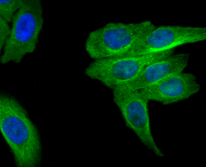
Fig1: ICC staining MRP2 in HepG2 cells (green). The nuclear counter stain is DAPI (blue). Cells were fixed in paraformaldehyde, permeabilised with 0.25% Triton X100/PBS.Application
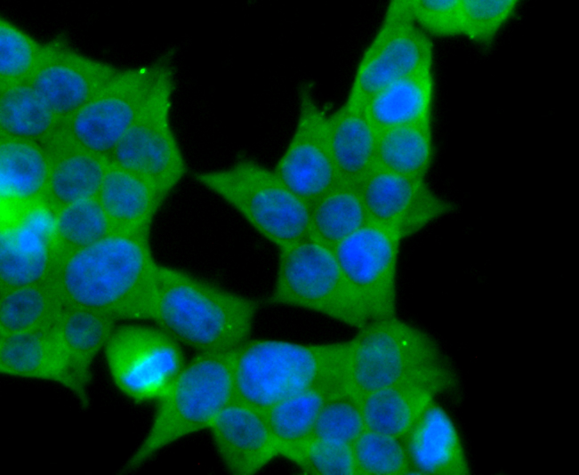
Fig2: ICC staining MRP2 in 293T cells (green). The nuclear counter stain is DAPI (blue). Cells were fixed in paraformaldehyde, permeabilised with 0.25% Triton X100/PBS.Application
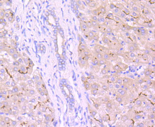
Fig3: Immunohistochemical analysis of paraffin-embedded human liver tissue using anti-MRP2 antibody. Counter stained with hematoxylin.Application
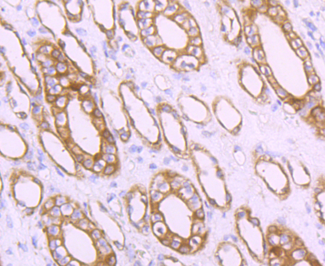
Fig4: Immunohistochemical analysis of paraffin-embedded human kidney tissue using anti-MRP2 antibody. Counter stained with hematoxylin.Application
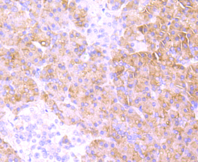
Fig5: Immunohistochemical analysis of paraffin-embedded human pancreas tissue using anti-MRP2 antibody. Counter stained with hematoxylin.Application
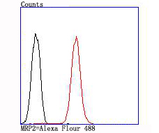
Fig6: Flow cytometric analysis of A549 cells with MRP2 antibody at 1/100 dilution (red) compared with an unlabelled control (cells without incubation with primary antibody; black).| Product Name | MRP2 [JA32-01] |
|---|---|
| Antibody Type | Primary Antibodies |
| Antigen Alias | ABC30 antibody abcC2 antibody ATP binding cassette sub family C (CFTR/MRP) member 2 antibody ATP binding cassette subfamily C member 2 antibody ATP-binding cassette sub-family C member 2 antibody Canalicular multidrug resistance protein antibody Canalicular multispecific organic anion transporter 1 antibody CMOAT antibody CMOAT1 antibody cMRP antibody DJS antibody KIAA1010 antibody MRP 2 antibody MRP2_HUMAN antibody Multidrug resistance associated protein 2 antibody Multidrug resistance-associated protein 2 antibody |
| Product description | Multi-drug resistance protein 2 (MRP2), also known as ABCC2, is an ATP binding cassette (ABC) transporter responsible for biliary excretion of xenobiotics, endobiotics, and their metabolites. Deficiency in ABCC2 results in the clinical disorder Dubin-Johnson syndrome. MRP2 is found to be expressed in a variety of human cancers, and is associated with resistance of tumor cells to various anticancer drugs including cisplatin. The predicted molecular weight of MRP2 is 174 kDa, while mature MRP2 usually has a slower migration around 190-250 kDa due to the glycosylation. |
| Immunogen | recombinant protein |
| Clonality | Monoclonal |
|---|---|
| Isotype | IgG |
| Host Species | Recombinant rabbit |
| Tested Applications | WBICCIHCFC |
| WB:1:500-1:1,000 ICC:1:50-1:200 IHC:1:50-1:200 FC:1:50-1:100 |
|
| Species Reactivity | Human |
| Concentration | 1mg/ml |
| Alternative Names | ABC30 antibody abcC2 antibody ATP binding cassette sub family C (CFTR/MRP) member 2 antibody ATP binding cassette subfamily C member 2 antibody ATP-binding cassette sub-family C member 2 antibody Canalicular multidrug resistance protein antibody Canalicular multispecific organic anion transporter 1 antibody CMOAT antibody CMOAT1 antibody cMRP antibody DJS antibody KIAA1010 antibody MRP 2 antibody MRP2_HUMAN antibody Multidrug resistance associated protein 2 antibody Multidrug resistance-associated protein 2 antibody |
|---|---|
| Molecular Weight(MW) | 174 kDa |
| Cellular Localization | Membrane. |
| SwissProt ID | Q92887 |
|---|
Application
Fig1: ICC staining MRP2 in HepG2 cells (green). The nuclear counter stain is DAPI (blue). Cells were fixed in paraformaldehyde, permeabilised with 0.25% Triton X100/PBS.
Application
Fig2: ICC staining MRP2 in 293T cells (green). The nuclear counter stain is DAPI (blue). Cells were fixed in paraformaldehyde, permeabilised with 0.25% Triton X100/PBS.
Application
Fig3: Immunohistochemical analysis of paraffin-embedded human liver tissue using anti-MRP2 antibody. Counter stained with hematoxylin.
Application
Fig4: Immunohistochemical analysis of paraffin-embedded human kidney tissue using anti-MRP2 antibody. Counter stained with hematoxylin.
Application
Fig5: Immunohistochemical analysis of paraffin-embedded human pancreas tissue using anti-MRP2 antibody. Counter stained with hematoxylin.
Application
Fig6: Flow cytometric analysis of A549 cells with MRP2 antibody at 1/100 dilution (red) compared with an unlabelled control (cells without incubation with primary antibody; black).| Positive Control | HepG2, A549, human liver tissue, human kidney tissue, human pancreas tissue. |
|---|---|
| Application Notes | WB:1:500-1:1,000 ICC:1:50-1:200 IHC:1:50-1:200 FC:1:50-1:100 |
| Form | Liquid |
|---|---|
| Storage Instructions | Store at +4℃ after thawing. Aliquot store at -20℃ or -80℃. Avoid repeated freeze / thaw cycles. |
| Storage Buffer | 1*TBS (pH7.4), 1%BSA, 40%Glycerol. Preservative: 0.05% Sodium Azide. |


