Application
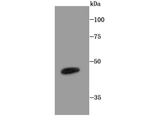
Fig1: Western blot analysis of LXR alpha on mouse colon tissue lysate using anti-LXR alpha antibody at 1/1,000 dilution.Application
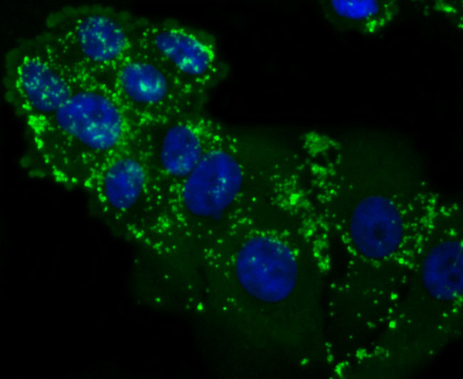
Fig2: ICC staining LXR alpha in HepG2 cells (green). The nuclear counter stain is DAPI (blue). Cells were fixed in paraformaldehyde, permeabilised with 0.25% Triton X100/PBS.Application
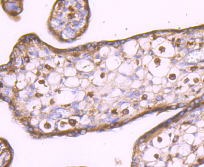
Fig3: Immunohistochemical analysis of paraffin-embedded human placenta tissue using anti-LXR alpha antibody. Counter stained with hematoxylin.Application
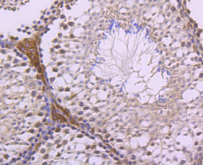
Fig4: Immunohistochemical analysis of paraffin-embedded mouse testes tissue using anti-LXR alpha antibody. Counter stained with hematoxylin.Application
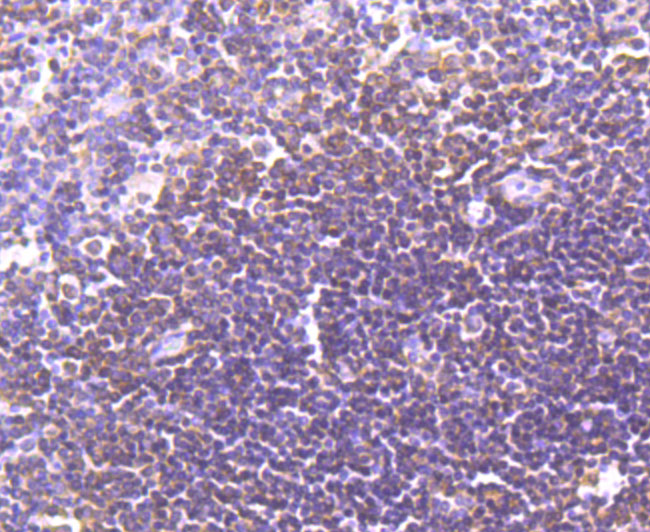
Fig5: Immunohistochemical analysis of paraffin-embedded human spleen tissue using anti-LXR alpha antibody. Counter stained with hematoxylin.Application
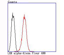
Fig6: Flow cytometric analysis of HepG2 cells with LXR alpha antibody at 1/100 dilution (red) compared with an unlabelled control (cells without incubation with primary antibody; black).| Product Name | LXR alpha [JA20-38] |
|---|---|
| Antibody Type | Primary Antibodies |
| Antigen Alias | Liver X receptor alpha antibody LXR a antibody LXRA antibody NR1H3 antibody NR1H3_HUMAN antibody Nuclear receptor subfamily 1 group H member 3 antibody Oxysterols receptor LXR alpha antibody Oxysterols receptor LXR-alpha antibody RLD 1 antibody RLD1 antibody |
| Product description | Orphan receptor. Interaction with RXR shifts RXR from its role as a silent DNA-binding partner to an active ligand-binding subunit in mediating retinoid responses through target genes defined by LXRES. LXRES are DR4-type response elements characterized by direct repeats of two similar hexanuclotide half-sites spaced by four nucleotides. Plays an important role in the regulation of cholesterol homeostasis, regulating cholesterol uptake through MYLIP-dependent ubiquitination of LDLR, VLDLR and LRP8. |
| Immunogen | recombinant protein |
| Clonality | Monoclonal |
|---|---|
| Isotype | IgG |
| Host Species | Recombinant rabbit |
| Tested Applications | WBICCIHCFC |
| WB:1:500 IHC:1:50-1:200 FC:1:50-1:100 |
|
| Species Reactivity | HumanMouseRat |
| Concentration | 1mg/ml |
| Alternative Names | Liver X receptor alpha antibody LXR a antibody LXRA antibody NR1H3 antibody NR1H3_HUMAN antibody Nuclear receptor subfamily 1 group H member 3 antibody Oxysterols receptor LXR alpha antibody Oxysterols receptor LXR-alpha antibody RLD 1 antibody RLD1 antibody |
|---|---|
| Molecular Weight(MW) | 50 kDa |
| Cellular Localization | Nucleus. |
Application
Fig1: Western blot analysis of LXR alpha on mouse colon tissue lysate using anti-LXR alpha antibody at 1/1,000 dilution.
Application
Fig2: ICC staining LXR alpha in HepG2 cells (green). The nuclear counter stain is DAPI (blue). Cells were fixed in paraformaldehyde, permeabilised with 0.25% Triton X100/PBS.
Application
Fig3: Immunohistochemical analysis of paraffin-embedded human placenta tissue using anti-LXR alpha antibody. Counter stained with hematoxylin.
Application
Fig4: Immunohistochemical analysis of paraffin-embedded mouse testes tissue using anti-LXR alpha antibody. Counter stained with hematoxylin.
Application
Fig5: Immunohistochemical analysis of paraffin-embedded human spleen tissue using anti-LXR alpha antibody. Counter stained with hematoxylin.
Application
Fig6: Flow cytometric analysis of HepG2 cells with LXR alpha antibody at 1/100 dilution (red) compared with an unlabelled control (cells without incubation with primary antibody; black).| Positive Control | Mouse colon tissue lysate, Human spleen tissue, human placenta tissue, mouse testes tissue, HepG2. |
|---|---|
| Application Notes | WB:1:500 IHC:1:50-1:200 FC:1:50-1:100 |
| Form | Liquid |
|---|---|
| Storage Instructions | Store at +4℃ after thawing. Aliquot store at -20℃ or -80℃. Avoid repeated freeze / thaw cycles. |
| Storage Buffer | 1*TBS (pH7.4), 1%BSA, 40%Glycerol. Preservative: 0.05% Sodium Azide. |


