Application
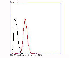
Fig3: Flow cytometric analysis of Hela cells with MRP1 antibody at 1/100 dilution (red) compared with an unlabelled control (cells without incubation with primary antibody; black).Application
Fig3: Flow cytometric analysis of Hela cells with MRP1 antibody at 1/100 dilution (red) compared with an unlabelled control (cells without incubation with primary antibody; black).Application
Fig3: Flow cytometric analysis of Hela cells with MRP1 antibody at 1/100 dilution (red) compared with an unlabelled control (cells without incubation with primary antibody; black).| Product Name | MRP1 [JA93-03] |
|---|---|
| Antibody Type | Primary Antibodies |
| Antigen Alias | ABC 29 antibody ABC29 antibody ABCC 1 antibody ABCC antibody Abcc1 antibody ATP binding cassette sub family C (CFTR/MRP) member 1 antibody ATP binding cassette sub-family C member 1 antibody ATP binding cassette subfamily C member 1 antibody ATP binding cassette transporter variant ABCC1delta ex13 antibody ATP binding cassette transporter variant ABCC1delta ex13&14 antibody ATP binding cassette transporter variant ABCC1delta ex25 antibody ATP binding cassette transporter variant ABCC1delta ex25&26 antibody ATP binding cassette, sub-family C (CFTR/MRP), member 1 antibody ATP-binding cassette sub-family C member 1 antibody DKFZp686N04233 antibody DKFZp781G125 antibody GS X antibody GSX antibody Leukotriene C(4) transporter antibody LTC4 transporter antibody MRP 1 antibody MRP antibody MRP1 antibody MRP1_HUMAN antibody Multidrug resistance associated protein 1 antibody Multidrug resistance protein antibody Multidrug resistance-associated protein 1 antibody Multiple drug resistance associated protein antibody Multiple drug resistance protein 1 antibody |
| Product description | The two members of the large family of ABC transporters known to confer multidrug resistance in human cancer cells are the Mdr-1 P-glycoprotein and the multidrug-resistance protein MRP1. MRP1 is an integral membrane protein that contains an Mdr-like core, an N-terminal membrane-bound region and a cytoplasmic linker, and it is expressed in various cerebral cells, as well as in lung, testis and peripheral blood. The MRP gene family also includes MRP2, which is alternatively designated cMOAT (for canalicular multispecific organic anion transporter) and MRP3, which are both conjugate export pumps expressed predominantly in hepatocytes. MRP2 localizes exclusively to the apical membrane and is constitutively expressed at a high level in normal liver cells. Conversely, MRP3 localizes to the basolateral membrane where it also mediates the transport of the organic anion S-(2,4-dinitrophenyl-) glutathione toward the basolateral side of the membrane. MRP3 is normally expressed at comparatively lower levels than MRP2 and increases only when secretion across the apical membrane by MRP2 is impaired. MRP6 protein is highly expressed in liver and kidney, whereas MRP4 and MRP5 are detected in various tissues yet at much lower levels of expression. |
| Immunogen | recombinant protein |
| Clonality | Monoclonal |
|---|---|
| Isotype | IgG |
| Host Species | Recombinant rabbit |
| Tested Applications | WBIHCFC |
| WB:1:500 IHC:1:50-1:200 |
|
| Species Reactivity | Human |
| Concentration | 1mg/ml |
| Alternative Names | ABC 29 antibody
ABC29 antibody
ABCC 1 antibody
ABCC antibody
Abcc1 antibody
ATP binding cassette sub family C (CFTR/MRP) member 1 antibody
ATP binding cassette sub-family C member 1 antibody
ATP binding cassette subfamily C member 1 antibody
ATP binding cassette transporter variant ABCC1delta ex13 antibody
ATP binding cassette transporter variant ABCC1delta ex13& 14 antibody ATP binding cassette transporter variant ABCC1delta ex25 antibody ATP binding cassette transporter variant ABCC1delta ex25& 26 antibody ATP binding cassette sub-family C (CFTR/MRP) member 1 antibody ATP-binding cassette sub-family C member 1 antibody DKFZp686N04233 antibody DKFZp781G125 antibody GS X antibody GSX antibody Leukotriene C(4) transporter antibody LTC4 transporter antibody MRP 1 antibody MRP antibody MRP1 antibody MRP1_HUMAN antibody Multidrug resistance associated protein 1 antibody Multidrug resistance protein antibody Multidrug resistance-associated protein 1 antibody Multiple drug resistance associated protein antibody Multiple drug resistance protein 1 antibody |
|---|---|
| Molecular Weight(MW) | 171 kDa |
| Cellular Localization | Cell membrane. |
| SwissProt ID | P33527 |
|---|
Application
Fig3: Flow cytometric analysis of Hela cells with MRP1 antibody at 1/100 dilution (red) compared with an unlabelled control (cells without incubation with primary antibody; black).
Application
Fig3: Flow cytometric analysis of Hela cells with MRP1 antibody at 1/100 dilution (red) compared with an unlabelled control (cells without incubation with primary antibody; black).
Application
Fig3: Flow cytometric analysis of Hela cells with MRP1 antibody at 1/100 dilution (red) compared with an unlabelled control (cells without incubation with primary antibody; black).| Positive Control | Human tonsil tissue, human pancreas tissue, Hela. |
|---|---|
| Application Notes | WB:1:500 IHC:1:50-1:200 |
| Form | Liquid |
|---|---|
| Storage Instructions | Store at +4℃ after thawing. Aliquot store at -20℃ or -80℃. Avoid repeated freeze / thaw cycles. |
| Storage Buffer | 1*TBS (pH7.4), 1%BSA, 40%Glycerol. Preservative: 0.05% Sodium Azide. |


