Application
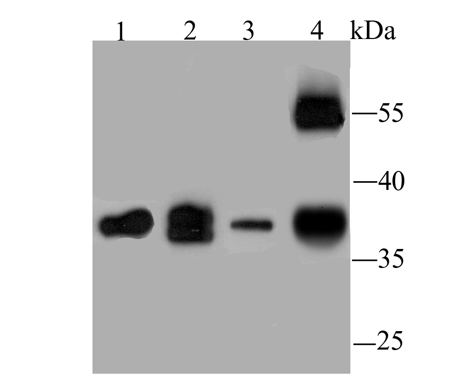
Fig1: Western blot analysis of WNT2 on different lysates using anti-WNT2 antibody at 1/1,000 dilution. Positive control: Lane 1: PC-3M Lane 2: Mouse brain tissue Lane 3: Jurkat Lane 4: Human lung tissueApplication
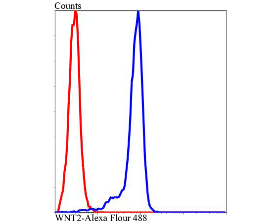
Fig2: Flow cytometric analysis of Jurkat cells with WNT2 antibody at 1/100 dilution (blue) compared with an unlabelled control (cells without incubation with primary antibody; red). Alexa Fluor 488-conjugated Goat anti rabbit IgG was used as the secondary antibody.| Product Name | WNT2 |
|---|---|
| Antibody Type | Primary Antibodies |
| Antigen Alias | Int 1 related protein antibody Int-1-like protein 1 antibody Int-1-related protein antibody INT1L 1 antibody INT1L1 antibody IRP antibody IRP protein antibody ONCOGENE INT1 LIKE 1 antibody Protein Wnt-2 antibody Secreted growth factor antibody Wingless type MMTV integration site family member 2 antibody WNT 2 antibody WNT2 antibody WNT2_HUMAN antibody |
| Product description | Belongs to the Wnt family. Ligand for members of the frizzled family of seven transmembrane receptors. Probable developmental protein. May be a signaling molecule which affects the development of discrete regions of tissues. Is likely to signal over only few cell diameters. |
| Immunogen | Peptide |
| Clonality | Polyclonal |
|---|---|
| Isotype | IgG |
| Host Species | Rabbit |
| Tested Applications | WBFC |
| WB:1:1,000-1:2,000 FC:1:100-1:200 |
|
| Species Reactivity | HumanMouse |
| Concentration | 1mg/ml |
| Alternative Names | Int 1 related protein antibody Int-1-like protein 1 antibody Int-1-related protein antibody INT1L 1 antibody INT1L1 antibody IRP antibody IRP protein antibody ONCOGENE INT1 LIKE 1 antibody Protein Wnt-2 antibody Secreted growth factor antibody Wingless type MMTV integration site family member 2 antibody WNT 2 antibody WNT2 antibody WNT2_HUMAN antibody |
|---|---|
| Molecular Weight(MW) | 37 kDa |
| Cellular Localization | Secreted. |
| SwissProt ID | P09544 |
|---|
Application
Fig1: Western blot analysis of WNT2 on different lysates using anti-WNT2 antibody at 1/1,000 dilution. Positive control: Lane 1: PC-3M Lane 2: Mouse brain tissue Lane 3: Jurkat Lane 4: Human lung tissue
Application
Fig2: Flow cytometric analysis of Jurkat cells with WNT2 antibody at 1/100 dilution (blue) compared with an unlabelled control (cells without incubation with primary antibody; red). Alexa Fluor 488-conjugated Goat anti rabbit IgG was used as the secondary antibody.| Positive Control | PC-3M, mouse brain tissue, Jurkat, human lung tissue. |
|---|---|
| Application Notes | WB:1:1,000-1:2,000 FC:1:100-1:200 |
| Form | Liquid |
|---|---|
| Storage Instructions | Store at +4℃ after thawing. Aliquot store at -20℃ or -80℃. Avoid repeated freeze / thaw cycles. |
| Storage Buffer | 1*TBS (pH7.4), 0.5%BSA, 50%Glycerol. Preservative: 0.05% Sodium Azide. |


