Application
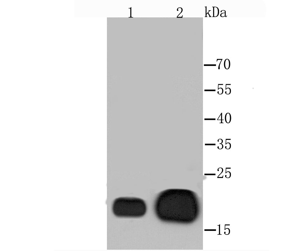
Fig1: Western blot analysis of Alpha B Crystallin on human skeleton musle (1) and mouse heart (2) tissues lysate using anti-Alpha B Crystallin antibody at 1/1,000 dilution.Application
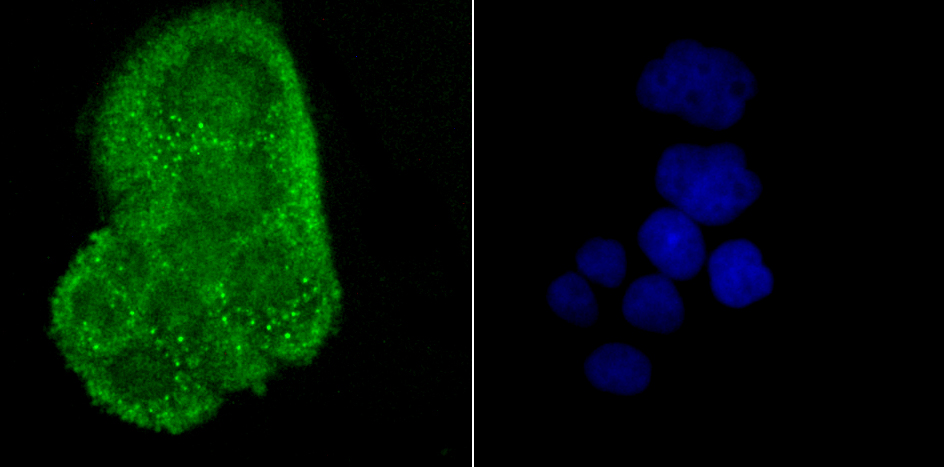
Fig2: ICC staining Alpha B Crystallin in Hela cells (green). The nuclear counter stain is DAPI (blue). Cells were fixed in paraformaldehyde, permeabilised with 0.25% Triton X100/PBS.Application
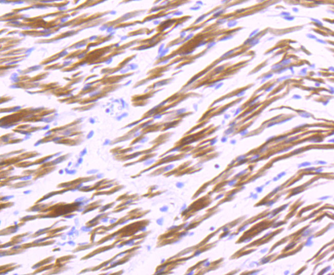
Fig3: Immunohistochemical analysis of paraffin-embedded human embryonic skeletal muscle tissue using anti-Alpha B Crystallin antibody. Counter stained with hematoxylin.Application
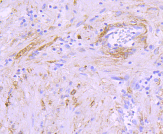
Fig4: Immunohistochemical analysis of paraffin-embedded human prostate tissue using anti-Alpha B Crystallin antibody. Counter stained with hematoxylin.| Product Name | Alpha B Crystallin [JA50-32] |
|---|---|
| Antibody Type | Primary Antibodies |
| Antigen Alias | AACRYA antibody Alpha B crystallin antibody Alpha crystallin B chain antibody Alpha(B)-crystallin antibody Alpha-crystallin B chain antibody CRYA2 antibody Cryab antibody CRYAB_HUMAN antibody Crystallin alpha B antibody Crystallin alpha polypeptide 2 antibody CTPP2 antibody Heat shock 20 kD like protein antibody Heat shock protein beta 5 antibody Heat shock protein beta-5 antibody HspB5 antibody Renal carcinoma antigen NY REN 27 antibody Renal carcinoma antigen NY-REN-27 antibody Rosenthal fiber component antibody |
| Product description | Crystallins are the major proteins of the vertebrate eye lens, where they maintain the transparency and refractive index of the lens. Crystallins are divided into α, β and γ families, and the β- and γ-crystallins also compose a superfamily. Crystallins usually contain seven distinct protein regions, inclu-ding four homologous motifs, a connecting peptide, and N- and C-terminal extensions. α-crystallins consist of three gene products, αA-, αB- and αC-crystallin, which are members of the small heat shock protein family (HSP 20). α-crystallins act as molecular chaperones by holding denatured proteins in large soluble aggregates. However, unlike other molecular chaperones, α-crystallins do not renature these proteins. Expression of αA-crystallin is restricted to the lens and defects of this gene cause the development of autosomal dominant congenital cataracts (ADCC). The human αB-crystallin gene product is expressed in many tissues, including lens, heart and skeletal muscle. Elevated expression of αB-crystallin is associated with many neurological diseases, and a missense mutation in this gene has co-segregated in a family with a Desmin-related myopathy. |
| Immunogen | recombinant protein |
| Clonality | Monoclonal |
|---|---|
| Isotype | IgG |
| Host Species | Recombinant rabbit |
| Tested Applications | WBIHCICC/IFIP |
| WB:1:500-1:2,000 ICC:1:50-1:200 IHC:1:50-1:200 IP:1:50 |
|
| Species Reactivity | HumanRat |
| Concentration | 1mg/ml |
| Alternative Names | AACRYA antibody Alpha B crystallin antibody Alpha crystallin B chain antibody Alpha(B)-crystallin antibody Alpha-crystallin B chain antibody CRYA2 antibody Cryab antibody CRYAB_HUMAN antibody Crystallin alpha B antibody Crystallin alpha polypeptide 2 antibody CTPP2 antibody Heat shock 20 kD like protein antibody Heat shock protein beta 5 antibody Heat shock protein beta-5 antibody HspB5 antibody Renal carcinoma antigen NY REN 27 antibody Renal carcinoma antigen NY-REN-27 antibody Rosenthal fiber component antibody |
|---|---|
| Molecular Weight(MW) | 20 kDa |
| Cellular Localization | Cytoplasm. Nucleus.? |
Application
Fig1: Western blot analysis of Alpha B Crystallin on human skeleton musle (1) and mouse heart (2) tissues lysate using anti-Alpha B Crystallin antibody at 1/1,000 dilution.
Application
Fig2: ICC staining Alpha B Crystallin in Hela cells (green). The nuclear counter stain is DAPI (blue). Cells were fixed in paraformaldehyde, permeabilised with 0.25% Triton X100/PBS.
Application
Fig3: Immunohistochemical analysis of paraffin-embedded human embryonic skeletal muscle tissue using anti-Alpha B Crystallin antibody. Counter stained with hematoxylin.
Application
Fig4: Immunohistochemical analysis of paraffin-embedded human prostate tissue using anti-Alpha B Crystallin antibody. Counter stained with hematoxylin.| Positive Control | Human skeleton musle, mouse heart tissues lysate, Hela, human embryonic skeletal muscle tissue, human prostate tissue. |
|---|---|
| Application Notes | WB:1:500-1:2,000 ICC:1:50-1:200 IHC:1:50-1:200 IP:1:50 |
| Form | Liquid |
|---|---|
| Storage Instructions | Store at +4℃ after thawing. Aliquot store at -20℃ or -80℃. Avoid repeated freeze / thaw cycles. |
| Storage Buffer | 1*TBS (pH7.4), 1%BSA, 40%Glycerol. Preservative: 0.05% Sodium Azide. |


