Application
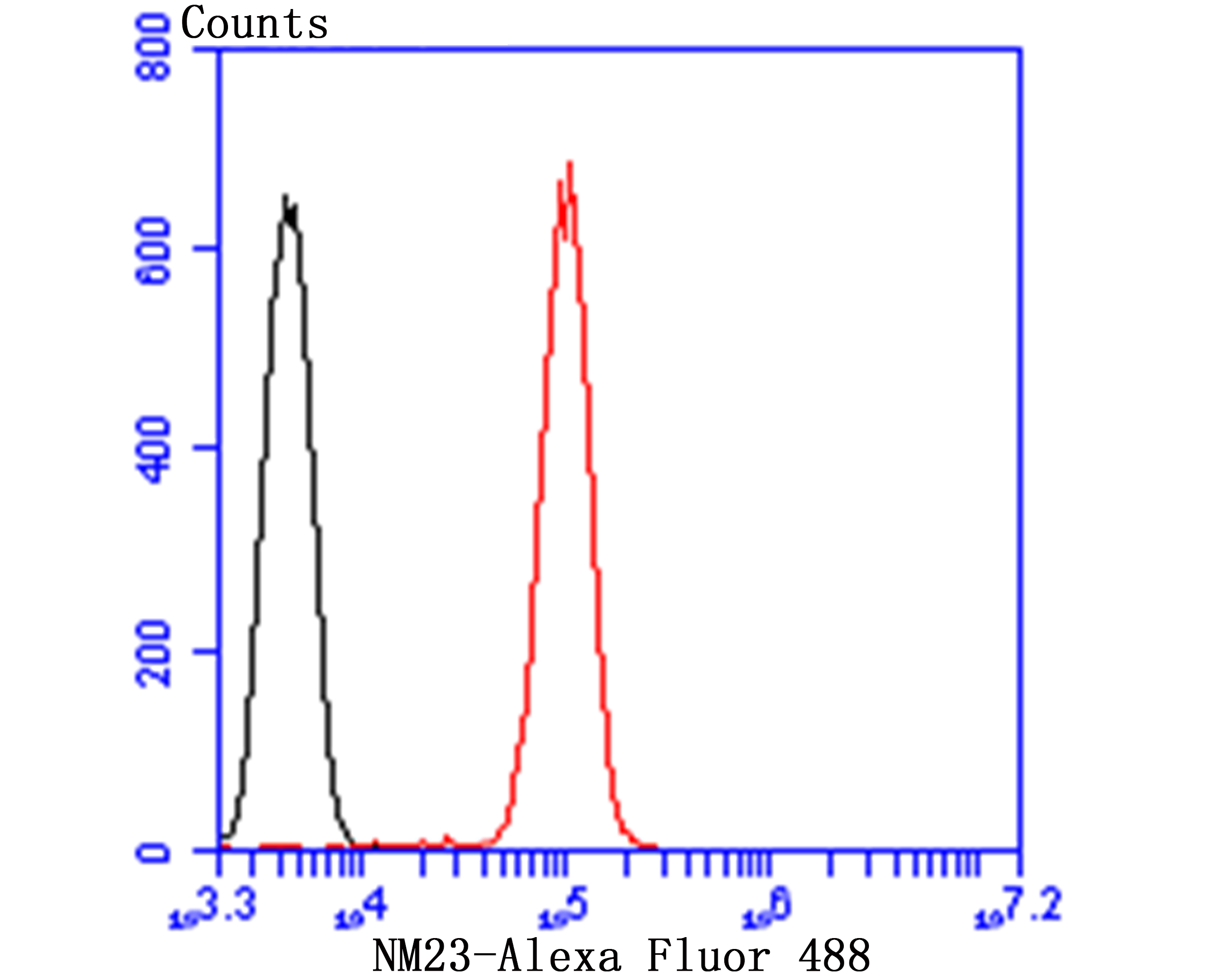
Fig7: Flow cytometric analysis of Jurkat cells with NM23 antibody at 1/100 dilution (red) compared with an unlabelled control (cells without incubation with primary antibody; black). Alexa Fluor 488-conjugated goat anti-rabbit IgG was used as the secondary antibody.Application
Fig7: Flow cytometric analysis of Jurkat cells with NM23 antibody at 1/100 dilution (red) compared with an unlabelled control (cells without incubation with primary antibody; black). Alexa Fluor 488-conjugated goat anti-rabbit IgG was used as the secondary antibody.Application
Fig7: Flow cytometric analysis of Jurkat cells with NM23 antibody at 1/100 dilution (red) compared with an unlabelled control (cells without incubation with primary antibody; black). Alexa Fluor 488-conjugated goat anti-rabbit IgG was used as the secondary antibody.Application
Fig7: Flow cytometric analysis of Jurkat cells with NM23 antibody at 1/100 dilution (red) compared with an unlabelled control (cells without incubation with primary antibody; black). Alexa Fluor 488-conjugated goat anti-rabbit IgG was used as the secondary antibody.Application
Fig7: Flow cytometric analysis of Jurkat cells with NM23 antibody at 1/100 dilution (red) compared with an unlabelled control (cells without incubation with primary antibody; black). Alexa Fluor 488-conjugated goat anti-rabbit IgG was used as the secondary antibody.Application
Fig7: Flow cytometric analysis of Jurkat cells with NM23 antibody at 1/100 dilution (red) compared with an unlabelled control (cells without incubation with primary antibody; black). Alexa Fluor 488-conjugated goat anti-rabbit IgG was used as the secondary antibody.Application
Fig7: Flow cytometric analysis of Jurkat cells with NM23 antibody at 1/100 dilution (red) compared with an unlabelled control (cells without incubation with primary antibody; black). Alexa Fluor 488-conjugated goat anti-rabbit IgG was used as the secondary antibody.| Product Name | NM23 |
|---|---|
| Antibody Type | Primary Antibodies |
| Antigen Alias | AWD antibody AWD, drosophila, homolog of antibody GAAD antibody Granzyme A activated DNase antibody Granzyme A-activated DNase antibody GZMA activated DNase antibody Metastasis inhibition factor NM23 antibody NB antibody NBS antibody NDK A antibody NDKA antibody NDKA_HUMAN antibody NDP kinase A antibody NDPK-A antibody NDPKA antibody NM23 antibody NM23 long variant, included antibody nm23-H1 antibody NM23-M1 antibody NM23H1B, included antibody NME/NM23 nucleoside diphosphate kinase 1 antibody Nme1 antibody NME1-NME2 spliced read-through transcript, included antibody Non-metastatic cells 1, protein (NM23A) expressed in antibody Nonmetastatic cells 1, protein expressed in antibody Nonmetastatic protein 23 antibody Nonmetastatic protein 23, homolog 1 antibody Nucleoside diphosphate kinase A antibody Tumor metastatic process-associated protein antibody |
| Product description | Major role in the synthesis of nucleoside triphosphates other than ATP. The ATP gamma phosphate is transferred to the NDP beta phosphate via a ping-pong mechanism, using a phosphorylated active-site intermediate. Possesses nucleoside-diphosphate kinase, serine/threonine-specific protein kinase, geranyl and farnesyl pyrophosphate kinase, histidine protein kinase and 3'-5' exonuclease activities. Involved in cell proliferation, differentiation and development, signal transduction, G protein-coupled receptor endocytosis, and gene expression. Required for neural development including neural patterning and cell fate determination. During GZMA-mediated cell death, works in concert with TREX1. NME1 nicks one strand of DNA and TREX1 removes bases from the free 3' end to enhance DNA damage and prevent DNA end reannealing and rapid repair. |
| Immunogen | Peptide |
| Clonality | Polyclonal |
|---|---|
| Isotype | IgG |
| Host Species | Rabbit |
| Tested Applications | ICCIHCFCWB |
| ICC:1:50-1:200 IHC:1:50-1:200 FC:1:50-1:100 WB:1:500 |
|
| Species Reactivity | HumanMouse |
| Concentration | 1mg/ml |
| Alternative Names | AWD antibody
AWD drosophila homolog of antibody GAAD antibody Granzyme A activated DNase antibody Granzyme A-activated DNase antibody GZMA activated DNase antibody Metastasis inhibition factor NM23 antibody NB antibody NBS antibody NDK A antibody NDKA antibody NDKA_HUMAN antibody NDP kinase A antibody NDPK-A antibody NDPKA antibody NM23 antibody NM23 long variant included antibody nm23-H1 antibody NM23-M1 antibody NM23H1B included antibody NME/NM23 nucleoside diphosphate kinase 1 antibody Nme1 antibody NME1-NME2 spliced read-through transcript included antibody Non-metastatic cells 1 protein (NM23A) expressed in antibody Nonmetastatic cells 1 protein expressed in antibody Nonmetastatic protein 23 antibody Nonmetastatic protein 23 homolog 1 antibody Nucleoside diphosphate kinase A antibody Tumor metastatic process-associated protein antibody |
|---|---|
| Molecular Weight(MW) | 17/19 kDa |
| Cellular Localization | Cytoplasm. Nucleus. |
Application
Fig7: Flow cytometric analysis of Jurkat cells with NM23 antibody at 1/100 dilution (red) compared with an unlabelled control (cells without incubation with primary antibody; black). Alexa Fluor 488-conjugated goat anti-rabbit IgG was used as the secondary antibody.
Application
Fig7: Flow cytometric analysis of Jurkat cells with NM23 antibody at 1/100 dilution (red) compared with an unlabelled control (cells without incubation with primary antibody; black). Alexa Fluor 488-conjugated goat anti-rabbit IgG was used as the secondary antibody.
Application
Fig7: Flow cytometric analysis of Jurkat cells with NM23 antibody at 1/100 dilution (red) compared with an unlabelled control (cells without incubation with primary antibody; black). Alexa Fluor 488-conjugated goat anti-rabbit IgG was used as the secondary antibody.
Application
Fig7: Flow cytometric analysis of Jurkat cells with NM23 antibody at 1/100 dilution (red) compared with an unlabelled control (cells without incubation with primary antibody; black). Alexa Fluor 488-conjugated goat anti-rabbit IgG was used as the secondary antibody.
Application
Fig7: Flow cytometric analysis of Jurkat cells with NM23 antibody at 1/100 dilution (red) compared with an unlabelled control (cells without incubation with primary antibody; black). Alexa Fluor 488-conjugated goat anti-rabbit IgG was used as the secondary antibody.
Application
Fig7: Flow cytometric analysis of Jurkat cells with NM23 antibody at 1/100 dilution (red) compared with an unlabelled control (cells without incubation with primary antibody; black). Alexa Fluor 488-conjugated goat anti-rabbit IgG was used as the secondary antibody.
Application
Fig7: Flow cytometric analysis of Jurkat cells with NM23 antibody at 1/100 dilution (red) compared with an unlabelled control (cells without incubation with primary antibody; black). Alexa Fluor 488-conjugated goat anti-rabbit IgG was used as the secondary antibody.| Positive Control | Hela, HepG2, MCF-7, human lung cancer tissue, human breast cancer tissue, mouse brain tissue. |
|---|---|
| Application Notes | ICC:1:50-1:200 IHC:1:50-1:200 FC:1:50-1:100 WB:1:500 |
| Form | Liquid |
|---|---|
| Storage Instructions | Store at +4℃ after thawing. Aliquot store at -20℃ or -80℃. Avoid repeated freeze / thaw cycles. |
| Storage Buffer | 1*TBS (pH7.4), 0.5%BSA, 50%Glycerol. Preservative: 0.05% Sodium Azide. |


