Application
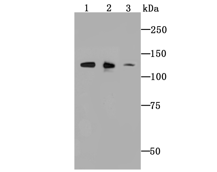
Fig1: Western blot analysis of ULK1 on different cell lysate using anti-ULK1 antibody at 1/1,000 dilution. Positive control: Lane1: MCF-7 Lane2: Mouse spleen Lane3: SH-SY5YApplication
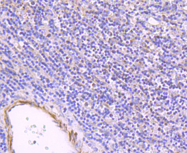
Fig2: Immunohistochemical analysis of paraffin-embedded human spleen tissue using anti-ULK1 antibody. Counter stained with hematoxylin.Application
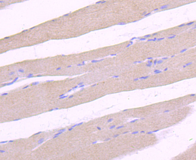
Fig3:Immunohistochemical analysis of paraffin-embedded rat skeleton muscle tissue using anti-ULK1 antibody. Counter stained with hematoxylin.Application
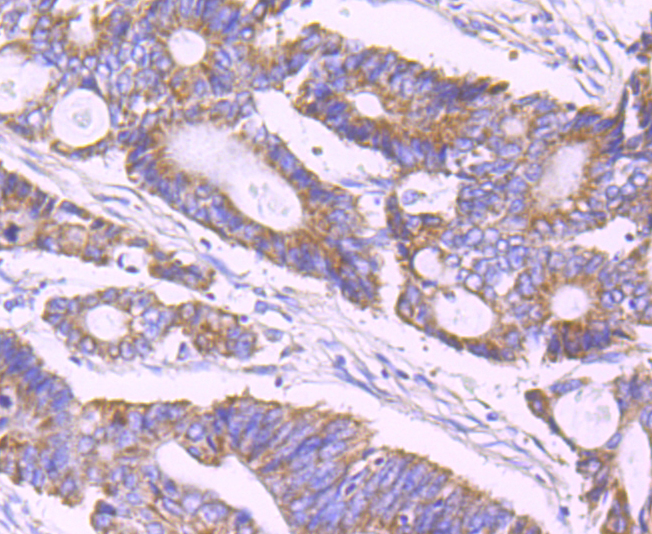
Fig4: Immunohistochemical analysis of paraffin-embedded human colon cancer tissue using anti-ULK1 antibody. Counter stained with hematoxylin.Application
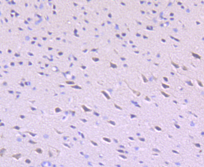
Fig5: Immunohistochemical analysis of paraffin-embedded mouse brain tissue using anti-ULK1 antibody. Counter stained with hematoxylin.| Product Name | ULK1 [JA58-36] |
|---|---|
| Antibody Type | Primary Antibodies |
| Antigen Alias | ATG 1 antibody ATG1 antibody ATG1 autophagy related 1 homolog antibody ATG1A antibody Autophagy related protein 1 homolog antibody Autophagy-related protein 1 homolog antibody FLJ38455 antibody FLJ46475 antibody hATG1 antibody KIAA0722 antibody Serine/threonine protein kinase ULK1 antibody Serine/threonine protein kinase Unc51.1 antibody Serine/threonine-protein kinase ULK1 antibody ULK 1 antibody ULK1 antibody ULK1_HUMAN antibody Unc 51 (C. elegans) like kinase 1 antibody UNC 51 antibody Unc 51 like kinase 1 antibody Unc-51 like kinase 1 (C. elegans) antibody Unc-51-like kinase 1 antibody UNC51 antibody UNC51, C. elegans, homolog of antibody Unc51.1 antibody |
| Product description | Serine/threonine-protein kinase involved in autophagy in response to starvation. Acts upstream of phosphatidylinositol 3-kinase PIK3C3 to regulate the formation of autophagophores, the precursors of autophagosomes. Part of regulatory feedback loops in autophagy: acts both as a downstream effector and negative regulator of mammalian target of rapamycin complex 1 (mTORC1) via interaction with RPTOR. Activated via phosphorylation by AMPK and also acts as a regulator of AMPK by mediating phosphorylation of AMPK subunits PRKAA1, PRKAB2 and PRKAG1, leading to negatively regulate AMPK activity. May phosphorylate ATG13/KIAA0652 and RPTOR; however such data need additional evidences. Plays a role early in neuronal differentiation and is required for granule cell axon formation. May also phosphorylate SESN2 and SQSTM1 to regulate autophagy. |
| Immunogen | recombinant protein |
| Clonality | Monoclonal |
|---|---|
| Isotype | IgG |
| Host Species | Recombinant rabbit |
| Tested Applications | WBIHC |
| WB:1:500-1:2,000 IHC:1:50-1:200 |
|
| Species Reactivity | HumanMouseRat |
| Concentration | 1mg/ml |
| Alternative Names | ATG 1 antibody
ATG1 antibody
ATG1 autophagy related 1 homolog antibody
ATG1A antibody
Autophagy related protein 1 homolog antibody
Autophagy-related protein 1 homolog antibody
FLJ38455 antibody
FLJ46475 antibody
hATG1 antibody
KIAA0722 antibody
Serine/threonine protein kinase ULK1 antibody
Serine/threonine protein kinase Unc51.1 antibody
Serine/threonine-protein kinase ULK1 antibody
ULK 1 antibody
ULK1 antibody
ULK1_HUMAN antibody
Unc 51 (C. elegans) like kinase 1 antibody
UNC 51 antibody
Unc 51 like kinase 1 antibody
Unc-51 like kinase 1 (C. elegans) antibody
Unc-51-like kinase 1 antibody
UNC51 antibody
UNC51 C. elegans homolog of antibody Unc51.1 antibody |
|---|---|
| Molecular Weight(MW) | 112 kDa |
| Cellular Localization | Cytoplasm. |
Application
Fig1: Western blot analysis of ULK1 on different cell lysate using anti-ULK1 antibody at 1/1,000 dilution. Positive control: Lane1: MCF-7 Lane2: Mouse spleen Lane3: SH-SY5Y
Application
Fig2: Immunohistochemical analysis of paraffin-embedded human spleen tissue using anti-ULK1 antibody. Counter stained with hematoxylin.
Application
Fig3:Immunohistochemical analysis of paraffin-embedded rat skeleton muscle tissue using anti-ULK1 antibody. Counter stained with hematoxylin.
Application
Fig4: Immunohistochemical analysis of paraffin-embedded human colon cancer tissue using anti-ULK1 antibody. Counter stained with hematoxylin.
Application
Fig5: Immunohistochemical analysis of paraffin-embedded mouse brain tissue using anti-ULK1 antibody. Counter stained with hematoxylin.| Positive Control | MCF-7, SH-SY5Y, mouse spleen tissue lysate, rat skeleton muscle tissue, human colon cancer tissue, mouse brain tissue, human spleen tissue. |
|---|---|
| Application Notes | WB:1:500-1:2,000 IHC:1:50-1:200 |
| Form | Liquid |
|---|---|
| Storage Instructions | Store at +4℃ after thawing. Aliquot store at -20℃ or -80℃. Avoid repeated freeze / thaw cycles. |
| Storage Buffer | 1*TBS (pH7.4), 1%BSA, 40%Glycerol. Preservative: 0.05% Sodium Azide. |


