Application
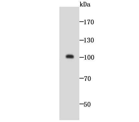
Fig1: Western blot analysis of GOLPH2 on SH-SY5Y cell lysate using anti-GOLPH2 antibody at 1/1,000 dilution.Application
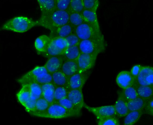
Fig2: ICC staining GOLPH2 in 293T cells (green). The nuclear counter stain is DAPI (blue). Cells were fixed in paraformaldehyde, permeabilised with 0.25% Triton X100/PBS.Application
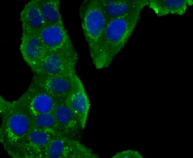
Fig3: ICC staining GOLPH2 in Hela cells (green). The nuclear counter stain is DAPI (blue). Cells were fixed in paraformaldehyde, permeabilised with 0.25% Triton X100/PBS.Application
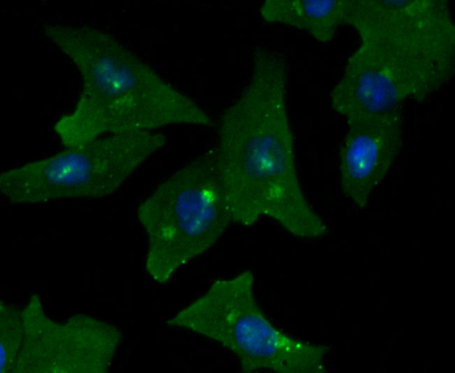
Fig4: ICC staining GOLPH2 in A549 cells (green). The nuclear counter stain is DAPI (blue). Cells were fixed in paraformaldehyde, permeabilised with 0.25% Triton X100/PBS.Application
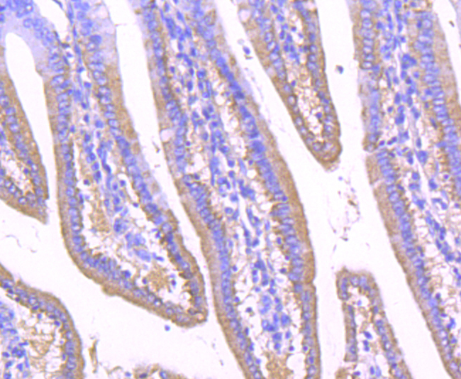
Fig5: Immunohistochemical analysis of paraffin-embedded mouse small intestine tissue using anti-GOLPH2 antibody. Counter stained with hematoxylin.Application
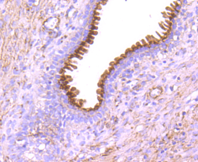
Fig6: Immunohistochemical analysis of paraffin-embedded human prostate tissue using anti-GOLPH2 antibody. Counter stained with hematoxylin.Application
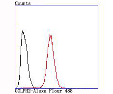
Fig7: Flow cytometric analysis of Hela cells with GOLPH2 antibody at 1/100 dilution (red) compared with an unlabelled control (cells without incubation with primary antibody; black).| Product Name | GOLPH2 [JA63-03] |
|---|---|
| Antibody Type | Primary Antibodies |
| Antigen Alias | bA379P1.3 antibody C9orf155 antibody Chromosome 9 open reading frame 155 antibody Golgi membrane protein 1 antibody Golgi membrane protein GP73 antibody Golgi phosphoprotein 2 antibody Golgi protein 73 kD antibody Golgi protein 73kD antibody GOLM 1 antibody GOLM1 antibody GOLM1_HUMAN antibody GOLPH 2 antibody GOLPH2 antibody GP 73 antibody GP73 antibody PSEC0257 antibody |
| Product description | GP73 (also known as Golgi phosphoprotein 2, GOLPH 2 or Golgi membrane protein), is a widely expressed, epithelial-specific, type II transmembrane protein which resides in the Golgi apparatus, where it is responsible for the posttranslational modification of proteins produced in the Rough ER while assisting in the transport of proteins through the Golgi. The human GP73 gene has been mapped within a BAC and localized to chromosome 9q21.33. GP73 levels rise in those who have been diagnosed with acute and chronic liver diseases. |
| Immunogen | recombinant protein |
| Clonality | Monoclonal |
|---|---|
| Isotype | IgG |
| Host Species | Recombinant rabbit |
| Tested Applications | WBICCIHCFC |
| WB:1:500-1:2,000 IHC:1:50 |
|
| Species Reactivity | HumanMouseRat |
| Concentration | 1mg/ml |
| Alternative Names | bA379P1.3 antibody C9orf155 antibody Chromosome 9 open reading frame 155 antibody Golgi membrane protein 1 antibody Golgi membrane protein GP73 antibody Golgi phosphoprotein 2 antibody Golgi protein 73 kD antibody Golgi protein 73kD antibody GOLM 1 antibody GOLM1 antibody GOLM1_HUMAN antibody GOLPH 2 antibody GOLPH2 antibody GP 73 antibody GP73 antibody PSEC0257 antibody |
|---|---|
| Molecular Weight(MW) | 100 kDa |
| Cellular Localization | Golgi apparatus. |
Application
Fig1: Western blot analysis of GOLPH2 on SH-SY5Y cell lysate using anti-GOLPH2 antibody at 1/1,000 dilution.
Application
Fig2: ICC staining GOLPH2 in 293T cells (green). The nuclear counter stain is DAPI (blue). Cells were fixed in paraformaldehyde, permeabilised with 0.25% Triton X100/PBS.
Application
Fig3: ICC staining GOLPH2 in Hela cells (green). The nuclear counter stain is DAPI (blue). Cells were fixed in paraformaldehyde, permeabilised with 0.25% Triton X100/PBS.
Application
Fig4: ICC staining GOLPH2 in A549 cells (green). The nuclear counter stain is DAPI (blue). Cells were fixed in paraformaldehyde, permeabilised with 0.25% Triton X100/PBS.
Application
Fig5: Immunohistochemical analysis of paraffin-embedded mouse small intestine tissue using anti-GOLPH2 antibody. Counter stained with hematoxylin.
Application
Fig6: Immunohistochemical analysis of paraffin-embedded human prostate tissue using anti-GOLPH2 antibody. Counter stained with hematoxylin.
Application
Fig7: Flow cytometric analysis of Hela cells with GOLPH2 antibody at 1/100 dilution (red) compared with an unlabelled control (cells without incubation with primary antibody; black).| Positive Control | SH-SY5Y cell lysate, 293T, A549, Hela, human prostate tissue, mouse small intestine tissue. |
|---|---|
| Application Notes | WB:1:500-1:2,000 IHC:1:50 |
| Form | Liquid |
|---|---|
| Storage Instructions | Store at +4℃ after thawing. Aliquot store at -20℃ or -80℃. Avoid repeated freeze / thaw cycles. |
| Storage Buffer | 1*TBS (pH7.4), 1%BSA, 40%Glycerol. Preservative: 0.05% Sodium Azide. |


