Application
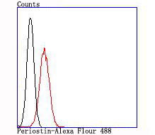
Fig2:Flow cytometric analysis of 293T cells with Periostin antibody at 1/100 dilution (red) compared with an unlabelled control (cells without incubation with primary antibody; black).Application
Fig2:Flow cytometric analysis of 293T cells with Periostin antibody at 1/100 dilution (red) compared with an unlabelled control (cells without incubation with primary antibody; black).| Product Name | Periostin [JA63-40] |
|---|---|
| Antibody Type | Primary Antibodies |
| Antigen Alias | Fasciclin-I like antibody MGC119510 antibody MGC119511 antibody OSF 2 antibody OSF-2 antibody OSF2 antibody Osteoblast specific factor 2 (fasciclin I like) antibody Osteoblast specific factor 2 antibody Osteoblast specific factor antibody Osteoblast-specific factor 2 antibody PDLPOSTN antibody Periodontal ligament specific periostin antibody Periostin antibody Periostin isoform thy2 antibody Periostin isoform thy4 antibody Periostin isoform thy6 antibody Periostin isoform thy8 antibody Periostin osteoblast specific factor antibody PN antibody POSTN antibody POSTN_HUMAN antibody RP11 412K4.1 antibody |
| Product description | Periostin (PN), also designated osteoblast-specific factor 2 (OSF-2), is a disulfide linked protein originally isolated as a osteoblast-specific factor. Periostin is a secreted protein that binds heparin and functions as a ligand for alpha(V)beta and alpha(V)beta integrins. In preosteoblasts, Periostin acts as a cell adhesion molecule and plays a role in osteoblast recruitment, spreading and attachment. Periostin is mainly detected in lower gastrointestinal tract, aorta, stomach, placenta, uterus and breast tissues but is up-regulated in epithelial ovarian tumors and overexpressed in breast cancer. Expression of Periostin is increased by bone morphogenetic protein (BMP2) and transforming growth factor β 1(TGF β 1). Periostin contains a typical signal sequence, followed by a cysteine-rich domain, a fourfold repeated domain, which shows homology with the insect protein fasciclin, and a C-terminal domain. |
| Immunogen | recombinant protein |
| Clonality | Monoclonal |
|---|---|
| Isotype | IgG |
| Host Species | Recombinant rabbit |
| Tested Applications | WBFC |
| WB:1:500-1:2,000 FC:1:50-1:100 |
|
| Species Reactivity | Human |
| Concentration | 1mg/ml |
| Alternative Names | Fasciclin-I like antibody MGC119510 antibody MGC119511 antibody OSF 2 antibody OSF-2 antibody OSF2 antibody Osteoblast specific factor 2 (fasciclin I like) antibody Osteoblast specific factor 2 antibody Osteoblast specific factor antibody Osteoblast-specific factor 2 antibody PDLPOSTN antibody Periodontal ligament specific periostin antibody Periostin antibody Periostin isoform thy2 antibody Periostin isoform thy4 antibody Periostin isoform thy6 antibody Periostin isoform thy8 antibody Periostin osteoblast specific factor antibody PN antibody POSTN antibody POSTN_HUMAN antibody RP11 412K4.1 antibody |
|---|---|
| Molecular Weight(MW) | 89 kDa |
| Cellular Localization | Secreted. |
| SwissProt ID | Q15063 |
|---|
Application
Fig2:Flow cytometric analysis of 293T cells with Periostin antibody at 1/100 dilution (red) compared with an unlabelled control (cells without incubation with primary antibody; black).
Application
Fig2:Flow cytometric analysis of 293T cells with Periostin antibody at 1/100 dilution (red) compared with an unlabelled control (cells without incubation with primary antibody; black).| Positive Control | Human placenta tissue lysate, 293T. |
|---|---|
| Application Notes | WB:1:500-1:2,000 FC:1:50-1:100 |
| Form | Liquid |
|---|---|
| Storage Instructions | Store at +4℃ after thawing. Aliquot store at -20℃ or -80℃. Avoid repeated freeze / thaw cycles. |
| Storage Buffer | 1*TBS (pH7.4), 1%BSA, 40%Glycerol. Preservative: 0.05% Sodium Azide. |


