Application
Fig9: Flow cytometric analysis of 293T cells with TGM6 antibody at 1/100 dilution (red) compared with an unlabelled control (cells without incubation with primary antibody; black). Alexa Fluor 488-conjugated Goat anti rabbit IgG was used as the secondary antibody.Application
Fig9: Flow cytometric analysis of 293T cells with TGM6 antibody at 1/100 dilution (red) compared with an unlabelled control (cells without incubation with primary antibody; black). Alexa Fluor 488-conjugated Goat anti rabbit IgG was used as the secondary antibody.Application
Fig9: Flow cytometric analysis of 293T cells with TGM6 antibody at 1/100 dilution (red) compared with an unlabelled control (cells without incubation with primary antibody; black). Alexa Fluor 488-conjugated Goat anti rabbit IgG was used as the secondary antibody.Application
Fig9: Flow cytometric analysis of 293T cells with TGM6 antibody at 1/100 dilution (red) compared with an unlabelled control (cells without incubation with primary antibody; black). Alexa Fluor 488-conjugated Goat anti rabbit IgG was used as the secondary antibody.Application
Fig9: Flow cytometric analysis of 293T cells with TGM6 antibody at 1/100 dilution (red) compared with an unlabelled control (cells without incubation with primary antibody; black). Alexa Fluor 488-conjugated Goat anti rabbit IgG was used as the secondary antibody.Application
Fig9: Flow cytometric analysis of 293T cells with TGM6 antibody at 1/100 dilution (red) compared with an unlabelled control (cells without incubation with primary antibody; black). Alexa Fluor 488-conjugated Goat anti rabbit IgG was used as the secondary antibody.Application
Fig9: Flow cytometric analysis of 293T cells with TGM6 antibody at 1/100 dilution (red) compared with an unlabelled control (cells without incubation with primary antibody; black). Alexa Fluor 488-conjugated Goat anti rabbit IgG was used as the secondary antibody.Application
Fig9: Flow cytometric analysis of 293T cells with TGM6 antibody at 1/100 dilution (red) compared with an unlabelled control (cells without incubation with primary antibody; black). Alexa Fluor 488-conjugated Goat anti rabbit IgG was used as the secondary antibody.Application
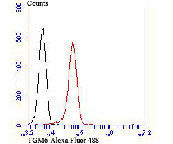
Fig9: Flow cytometric analysis of 293T cells with TGM6 antibody at 1/100 dilution (red) compared with an unlabelled control (cells without incubation with primary antibody; black). Alexa Fluor 488-conjugated Goat anti rabbit IgG was used as the secondary antibody.| Product Name | TGM6 |
|---|---|
| Antibody Type | Primary Antibodies |
| Antigen Alias | Protein-glutamine gamma-glutamyltransferase 6 antibody TG6 antibody TGase Y antibody TGase-3-like antibody TGase-6 antibody TGM3L_HUMAN antibody TGM6 antibody TGY antibody Transglutaminase Y antibody Transglutaminase-3-like antibody Transglutaminase-6 antibody |
| Product description | Transglutaminase 6 is a protein that in humans is encoded by the TGM6 gene. The protein encoded by this gene belongs to the transglutaminase superfamily. It catalyzes the cross-linking of proteins and the conjugation of polyamines to proteins. Mutations in this gene are associated with spinocerebellar ataxia type 35 (SCA35). Alternatively spliced transcript variants encoding different isoforms have been found for this gene. |
| Immunogen | Synthetic peptide corresponding to N-terminal of Mouse TGM6. |
| Clonality | Polyclonal |
|---|---|
| Isotype | IgG |
| Host Species | Rabbit |
| Tested Applications | WBICCIHCFC |
| WB:1:500: ICC:1:50-1:200 IHC:1:50-1:200 FC:1:50-1:100 |
|
| Species Reactivity | HumanMouseRat |
| Concentration | 1mg/ml |
| Alternative Names | Protein-glutamine gamma-glutamyltransferase 6 antibody TG6 antibody TGase Y antibody TGase-3-like antibody TGase-6 antibody TGM3L_HUMAN antibody TGM6 antibody TGY antibody Transglutaminase Y antibody Transglutaminase-3-like antibody Transglutaminase-6 antibody |
|---|---|
| Molecular Weight(MW) | 79/71 kDa |
| Cellular Localization | Cytoplasm. |
Application
Fig9: Flow cytometric analysis of 293T cells with TGM6 antibody at 1/100 dilution (red) compared with an unlabelled control (cells without incubation with primary antibody; black). Alexa Fluor 488-conjugated Goat anti rabbit IgG was used as the secondary antibody.
Application
Fig9: Flow cytometric analysis of 293T cells with TGM6 antibody at 1/100 dilution (red) compared with an unlabelled control (cells without incubation with primary antibody; black). Alexa Fluor 488-conjugated Goat anti rabbit IgG was used as the secondary antibody.
Application
Fig9: Flow cytometric analysis of 293T cells with TGM6 antibody at 1/100 dilution (red) compared with an unlabelled control (cells without incubation with primary antibody; black). Alexa Fluor 488-conjugated Goat anti rabbit IgG was used as the secondary antibody.
Application
Fig9: Flow cytometric analysis of 293T cells with TGM6 antibody at 1/100 dilution (red) compared with an unlabelled control (cells without incubation with primary antibody; black). Alexa Fluor 488-conjugated Goat anti rabbit IgG was used as the secondary antibody.
Application
Fig9: Flow cytometric analysis of 293T cells with TGM6 antibody at 1/100 dilution (red) compared with an unlabelled control (cells without incubation with primary antibody; black). Alexa Fluor 488-conjugated Goat anti rabbit IgG was used as the secondary antibody.
Application
Fig9: Flow cytometric analysis of 293T cells with TGM6 antibody at 1/100 dilution (red) compared with an unlabelled control (cells without incubation with primary antibody; black). Alexa Fluor 488-conjugated Goat anti rabbit IgG was used as the secondary antibody.
Application
Fig9: Flow cytometric analysis of 293T cells with TGM6 antibody at 1/100 dilution (red) compared with an unlabelled control (cells without incubation with primary antibody; black). Alexa Fluor 488-conjugated Goat anti rabbit IgG was used as the secondary antibody.
Application
Fig9: Flow cytometric analysis of 293T cells with TGM6 antibody at 1/100 dilution (red) compared with an unlabelled control (cells without incubation with primary antibody; black). Alexa Fluor 488-conjugated Goat anti rabbit IgG was used as the secondary antibody.
Application
Fig9: Flow cytometric analysis of 293T cells with TGM6 antibody at 1/100 dilution (red) compared with an unlabelled control (cells without incubation with primary antibody; black). Alexa Fluor 488-conjugated Goat anti rabbit IgG was used as the secondary antibody.| Positive Control | 293T, A549, SH-SY5Y, rat brain tissue, human kidney tissue, mouse testis tissue, mouse brain tissue, mouse spleen tissue. |
|---|---|
| Application Notes | WB:1:500: ICC:1:50-1:200 IHC:1:50-1:200 FC:1:50-1:100 |
| Form | Liquid |
|---|---|
| Storage Instructions | Store at +4℃ after thawing. Aliquot store at -20℃ or -80℃. Avoid repeated freeze / thaw cycles. |
| Storage Buffer | 1*TBS (pH7.4), 0.5%BSA, 50%Glycerol. Preservative: 0.05% Sodium Azide. |


