Application
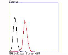
Fig6: Flow cytometric analysis of NIH-3T3 cells with PDK1 antibody at 1/100 dilution (red) compared with an unlabelled control (cells without incubation with primary antibody; black).Application
Fig6: Flow cytometric analysis of NIH-3T3 cells with PDK1 antibody at 1/100 dilution (red) compared with an unlabelled control (cells without incubation with primary antibody; black).Application
Fig6: Flow cytometric analysis of NIH-3T3 cells with PDK1 antibody at 1/100 dilution (red) compared with an unlabelled control (cells without incubation with primary antibody; black).Application
Fig6: Flow cytometric analysis of NIH-3T3 cells with PDK1 antibody at 1/100 dilution (red) compared with an unlabelled control (cells without incubation with primary antibody; black).Application
Fig6: Flow cytometric analysis of NIH-3T3 cells with PDK1 antibody at 1/100 dilution (red) compared with an unlabelled control (cells without incubation with primary antibody; black).Application
Fig6: Flow cytometric analysis of NIH-3T3 cells with PDK1 antibody at 1/100 dilution (red) compared with an unlabelled control (cells without incubation with primary antibody; black).| Product Name | PDK1 [JA67-30] |
|---|---|
| Antibody Type | Primary Antibodies |
| Product description | Mitochondrial pyruvate dehydrogenase (PDH) catalyzes the oxidative decarboxylation of pyruvate and plays a central role in the regulation of homeostasis of carbohydrate fuels in mammals. PDH activity is controlled by a phosphorylation/dephosphorylation cycle, phosphorylation leading to inactivation and dephosphorylation leading to reactivation of PDH. The phosphorylation of PDH is catalyzed by pyruvate dehydrogenase kinase (PDK), the activity of which is stimulated by the products of PDH catalysis. PDK1 consists of alpha and beta subunits; the kinase activity resides in the alpha subunit. Three PDK isoenzymes have been identified in humans (PDK1, 2 and 3) and two have been identified in rodent (PDK1 and 2). |
| Immunogen | recombinant protein |
| Clonality | Monoclonal |
|---|---|
| Isotype | IgG |
| Host Species | Recombinant rabbit |
| Tested Applications | WBIHCIPFC |
| WB:1:500-1:2,000 ICC:1:50-1:200: IHC:1:50-1:200 FC:1:50-1:100 |
|
| Species Reactivity | HumanMouseRat |
| Concentration | 1mg/ml |
| Molecular Weight(MW) | 49 kDa |
|---|---|
| Cellular Localization | Mitochondrion matrix. Nucleus. |
Application
Fig6: Flow cytometric analysis of NIH-3T3 cells with PDK1 antibody at 1/100 dilution (red) compared with an unlabelled control (cells without incubation with primary antibody; black).
Application
Fig6: Flow cytometric analysis of NIH-3T3 cells with PDK1 antibody at 1/100 dilution (red) compared with an unlabelled control (cells without incubation with primary antibody; black).
Application
Fig6: Flow cytometric analysis of NIH-3T3 cells with PDK1 antibody at 1/100 dilution (red) compared with an unlabelled control (cells without incubation with primary antibody; black).
Application
Fig6: Flow cytometric analysis of NIH-3T3 cells with PDK1 antibody at 1/100 dilution (red) compared with an unlabelled control (cells without incubation with primary antibody; black).
Application
Fig6: Flow cytometric analysis of NIH-3T3 cells with PDK1 antibody at 1/100 dilution (red) compared with an unlabelled control (cells without incubation with primary antibody; black).
Application
Fig6: Flow cytometric analysis of NIH-3T3 cells with PDK1 antibody at 1/100 dilution (red) compared with an unlabelled control (cells without incubation with primary antibody; black).| Positive Control | Rat heart tissue lysate, Hela, NIH-3T3, human kidney tissue, mouse heart tissue. |
|---|---|
| Application Notes | WB:1:500-1:2,000 ICC:1:50-1:200: IHC:1:50-1:200 FC:1:50-1:100 |
| Form | Liquid |
|---|---|
| Storage Instructions | Store at +4℃ after thawing. Aliquot store at -20℃ or -80℃. Avoid repeated freeze / thaw cycles. |
| Storage Buffer | 1*TBS (pH7.4), 1%BSA, 40%Glycerol. Preservative: 0.05% Sodium Azide. |


