Application
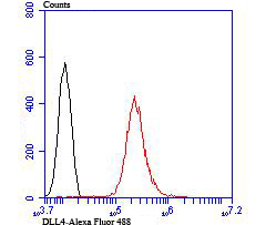
Fig7: Flow cytometric analysis of HUVEC cells with DLL4 antibody at 1/100 dilution (red) compared with an unlabelled control (cells without incubation with primary antibody; black).Application
Fig7: Flow cytometric analysis of HUVEC cells with DLL4 antibody at 1/100 dilution (red) compared with an unlabelled control (cells without incubation with primary antibody; black).Application
Fig7: Flow cytometric analysis of HUVEC cells with DLL4 antibody at 1/100 dilution (red) compared with an unlabelled control (cells without incubation with primary antibody; black).Application
Fig7: Flow cytometric analysis of HUVEC cells with DLL4 antibody at 1/100 dilution (red) compared with an unlabelled control (cells without incubation with primary antibody; black).Application
Fig7: Flow cytometric analysis of HUVEC cells with DLL4 antibody at 1/100 dilution (red) compared with an unlabelled control (cells without incubation with primary antibody; black).Application
Fig7: Flow cytometric analysis of HUVEC cells with DLL4 antibody at 1/100 dilution (red) compared with an unlabelled control (cells without incubation with primary antibody; black).Application
Fig7: Flow cytometric analysis of HUVEC cells with DLL4 antibody at 1/100 dilution (red) compared with an unlabelled control (cells without incubation with primary antibody; black).| Product Name | DLL4 |
|---|---|
| Antibody Type | Primary Antibodies |
| Antigen Alias | AOS6 antibody Delta 4 antibody delta 4 precursor antibody Delta ligand 4 antibody delta ligand 4 precursor antibody Delta like 4 antibody Delta like 4 homolog antibody Delta like 4 protein antibody Delta like canonical Notch ligand 4 antibody Delta like protein 4 antibody Delta-like 4 (Drosophila) antibody Delta-like protein 4 antibody Delta4 antibody DLL 4 antibody Dll4 antibody DLL4_HUMAN antibody Drosophila Delta homolog 4 antibody hdelta2 antibody Homeobox protein DLL-4 antibody MGC126344 antibody Notch ligand delta 2 antibody Notch ligand DLL4 antibody Notch ligand DLL4 precursor antibody XDLL-4 antibody |
| Product description | The LIN-12/Notch family of transmembrane receptors is believed to play a central role in development by regulating cell fate decisions. Notch proteins have been found to be overexpressed or rearranged in human tumors. Ligands for Notch include Jagged, Jagged-2 and Delta. While blocking the differentiation of progenitor cells into the B-cell lineage, Delta promotes the emergence of a population of cells with T cell/NK-cell characteristics. The protein is a membrane protein expressed in heart, pancreas, brain and muscle during gastrulation and early organogenesis and in adult heart and lung. Delta-4 is a membrane protein that activates Notch-1 and Notch-4. It is expressed in a wide range of adult and fetal tissues, especially in vascular endothelium. |
| Immunogen | Recombinant protein |
| Clonality | Polyclonal |
|---|---|
| Isotype | IgG |
| Host Species | Rabbit |
| Tested Applications | WBICCIHCFC |
| WB:1:500-1:1,000 ICC:1:50-1:200 IHC:1:50-1:200 FC:1:50-1:100 |
|
| Species Reactivity | HumanMouse |
| Concentration | 1mg/ml |
| Alternative Names | AOS6 antibody Delta 4 antibody delta 4 precursor antibody Delta ligand 4 antibody delta ligand 4 precursor antibody Delta like 4 antibody Delta like 4 homolog antibody Delta like 4 protein antibody Delta like canonical Notch ligand 4 antibody Delta like protein 4 antibody Delta-like 4 (Drosophila) antibody Delta-like protein 4 antibody Delta4 antibody DLL 4 antibody Dll4 antibody DLL4_HUMAN antibody Drosophila Delta homolog 4 antibody hdelta2 antibody Homeobox protein DLL-4 antibody MGC126344 antibody Notch ligand delta 2 antibody Notch ligand DLL4 antibody Notch ligand DLL4 precursor antibody XDLL-4 antibody |
|---|---|
| Molecular Weight(MW) | 74 kDa (Predicted band size) |
| Cellular Localization | Cell membrane. |
Application
Fig7: Flow cytometric analysis of HUVEC cells with DLL4 antibody at 1/100 dilution (red) compared with an unlabelled control (cells without incubation with primary antibody; black).
Application
Fig7: Flow cytometric analysis of HUVEC cells with DLL4 antibody at 1/100 dilution (red) compared with an unlabelled control (cells without incubation with primary antibody; black).
Application
Fig7: Flow cytometric analysis of HUVEC cells with DLL4 antibody at 1/100 dilution (red) compared with an unlabelled control (cells without incubation with primary antibody; black).
Application
Fig7: Flow cytometric analysis of HUVEC cells with DLL4 antibody at 1/100 dilution (red) compared with an unlabelled control (cells without incubation with primary antibody; black).
Application
Fig7: Flow cytometric analysis of HUVEC cells with DLL4 antibody at 1/100 dilution (red) compared with an unlabelled control (cells without incubation with primary antibody; black).
Application
Fig7: Flow cytometric analysis of HUVEC cells with DLL4 antibody at 1/100 dilution (red) compared with an unlabelled control (cells without incubation with primary antibody; black).
Application
Fig7: Flow cytometric analysis of HUVEC cells with DLL4 antibody at 1/100 dilution (red) compared with an unlabelled control (cells without incubation with primary antibody; black).| Positive Control | Mouse placenta tissue lysate, HUVEC, 293T, PMVEC, human liver tissue, human kidney tissue. |
|---|---|
| Application Notes | WB:1:500-1:1,000 ICC:1:50-1:200 IHC:1:50-1:200 FC:1:50-1:100 |
| Form | Liquid |
|---|---|
| Storage Instructions | Store at +4℃ after thawing. Aliquot store at -20℃ or -80℃. Avoid repeated freeze / thaw cycles. |
| Storage Buffer | 1*TBS (pH7.4), 0.5%BSA, 50%Glycerol. Preservative: 0.05% Sodium Azide. |


