WB
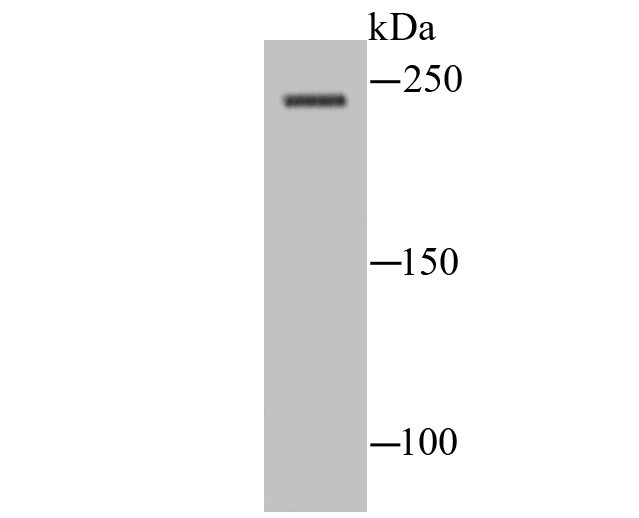
Western blot analysis of NaV1.8 on rat spinal cord lysate using anti-NaV1.8 antibody at 1/100 dilution.WB
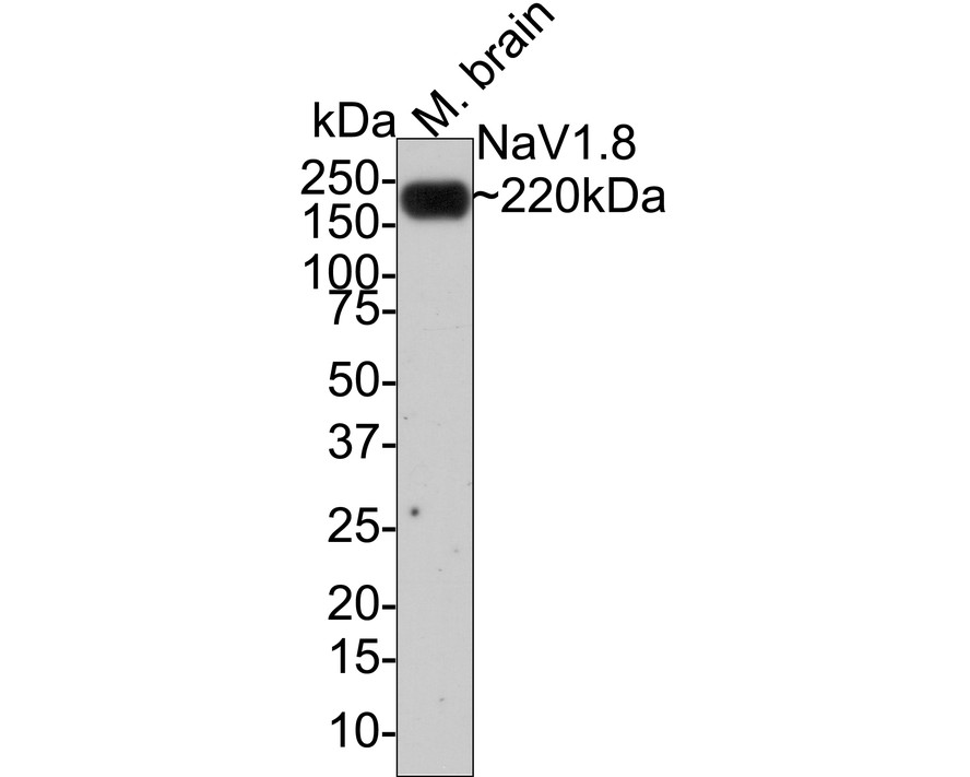
Western blot analysis of NaV1.8 on mouse brain tissue lysates with Rabbit anti-NaV1.8 antibody at 1/1,000 dilution. Lysates/proteins at 40 µg/Lane. Exposure time: 1 minute; 4-20% SDS-PAGE gel. Proteins were transferred to a PVDF membrane and blocked with 5% NFDM/TBST for 1 hour at room temperature. The primary antibody at 1/1,000 dilution was used in 5% NFDM/TBST at room temperature for 2 hours. Goat Anti-Rabbit IgG - HRP Secondary Antibody at 1:100,000 dilution was used for 1 hour at room temperature.IHC
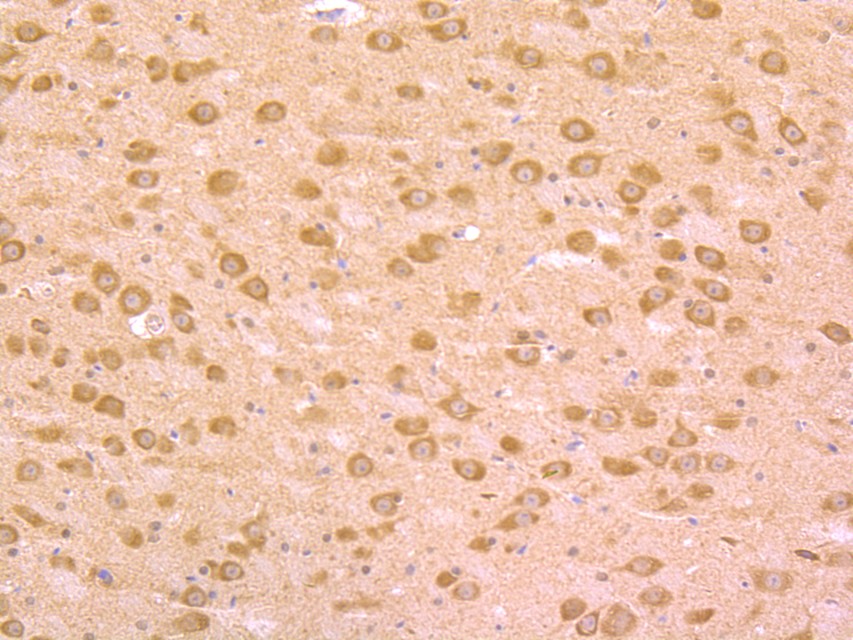
Immunohistochemical analysis of paraffin-embedded mouse brain tissue using anti-NaV1.8 antibody. The section was pre-treated using heat mediated antigen retrieval with Tris-EDTA buffer (pH 9.0) for 20 minutes.The tissues were blocked in 1% BSA for 30 minutes at room temperature, washed with ddH2O and PBS, and then probed with the primary antibody (1/50) for 30 minutes at room temperature. The detection was performed using an HRP conjugated compact polymer system. DAB was used as the chromogen. Tissues were counterstained with hematoxylin and mounted with DPX.| Product Name | NaV1.8 Rabbit Polyclonal Antibody |
|---|---|
| Antibody Type | Primary Antibodies |
| Product description | Tetrodotoxin-resistant channel that mediates the voltage-dependent sodium ion permeability of excitable membranes. Assuming opened or closed conformations in response to the voltage difference across the membrane, the protein forms a sodium-selective channel through which sodium ions may pass in accordance with their electrochemical gradient. Plays a role in neuropathic pain mechanisms. |
| Immunogen | Synthetic peptide within rat NaV18 aa 1,907-1,956 / 1,956. |
| Clonality | Polyclonal |
|---|---|
| Isotype | IgG |
| Host Species | Rabbit |
| Tested Applications | IHCWB |
| WB:1:100-1:1000 IHC:1:50 |
|
| Species Reactivity | MouseRat |
| Concentration | 1mg/ml |
| Purification | Affinity purified |
| Alternative Names | hPN3 antibody mPN3 antibody Peripheral nerve sodium channel 3 antibody Pn3 (gene name) antibody PN3 antibody SCN10A antibody SCNAA_HUMAN antibody Sensory neuron sodium channel antibody Sns (gene name) antibody SNS antibody Sodium channel protein type 10 subunit alpha antibody Sodium channel protein type X alpha subunit antibody Sodium channel protein type X subunit alpha antibody Voltage-gated sodium channel alpha subunit Nav1.8 antibody Voltage-gated sodium channel subunit alpha Nav1.8 antibody |
|---|---|
| Molecular Weight(MW) | 220 kDa |
| Cellular Localization | Plasma membrane. |
WB
Western blot analysis of NaV1.8 on rat spinal cord lysate using anti-NaV1.8 antibody at 1/100 dilution.
WB
Western blot analysis of NaV1.8 on mouse brain tissue lysates with Rabbit anti-NaV1.8 antibody at 1/1,000 dilution. Lysates/proteins at 40 µg/Lane. Exposure time: 1 minute; 4-20% SDS-PAGE gel. Proteins were transferred to a PVDF membrane and blocked with 5% NFDM/TBST for 1 hour at room temperature. The primary antibody at 1/1,000 dilution was used in 5% NFDM/TBST at room temperature for 2 hours. Goat Anti-Rabbit IgG - HRP Secondary Antibody at 1:100,000 dilution was used for 1 hour at room temperature.
IHC
Immunohistochemical analysis of paraffin-embedded mouse brain tissue using anti-NaV1.8 antibody. The section was pre-treated using heat mediated antigen retrieval with Tris-EDTA buffer (pH 9.0) for 20 minutes.The tissues were blocked in 1% BSA for 30 minutes at room temperature, washed with ddH2O and PBS, and then probed with the primary antibody (1/50) for 30 minutes at room temperature. The detection was performed using an HRP conjugated compact polymer system. DAB was used as the chromogen. Tissues were counterstained with hematoxylin and mounted with DPX.| Positive Control | Rat spinal cord lysate, mouse brain tissue. |
|---|---|
| Application Notes | WB:1:100-1:1000 IHC:1:50 |
| Form | Liquid |
|---|---|
| Storage Instructions | Store at +4℃ after thawing. Aliquot store at -20℃ or -80℃. Avoid repeated freeze / thaw cycles. |
| Storage Buffer | 1*TBS (pH7.4), 0.5%BSA, 50%Glycerol. Preservative: 0.05% Sodium Azide. |


