Application
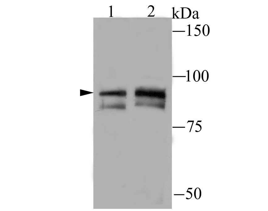
Fig1: Western blot analysis of TNFAIP3 on Jurkat (1) and Daudi (2) cell lysate using anti-TNFAIP3 antibody at 1/100 dilution.Application
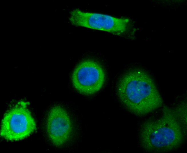
Fig2: ICC staining TNFAIP3 in A431 cells (green). The nuclear counter stain is DAPI (blue). Cells were fixed in paraformaldehyde, permeabilised with 0.25% Triton X100/PBS.Application
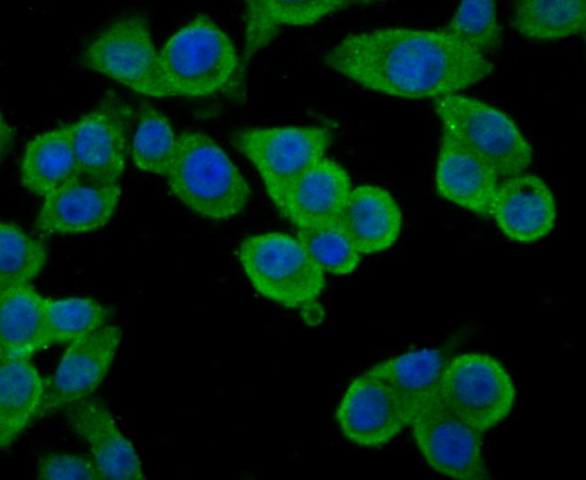
Fig3: ICC staining TNFAIP3 in LOVO cells (green). The nuclear counter stain is DAPI (blue). Cells were fixed in paraformaldehyde, permeabilised with 0.25% Triton X100/PBS.Application
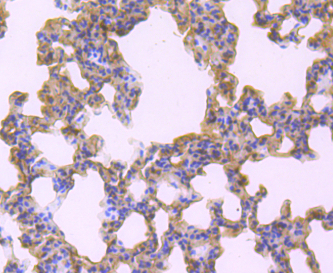
Fig4: Immunohistochemical analysis of paraffin-embedded rat lung tissue using anti-TNFAIP3 antibody. Counter stained with hematoxylin.Application
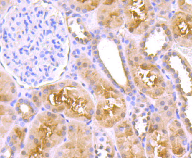
Fig5: Immunohistochemical analysis of paraffin-embedded human kidney tissue using anti-TNFAIP3 antibody. Counter stained with hematoxylin.Application
Fig6: Immunohistochemical analysis of paraffin-embedded human kidney tissue using anti-TNFAIP3 antibody. Counter stained with hematoxylin.Application
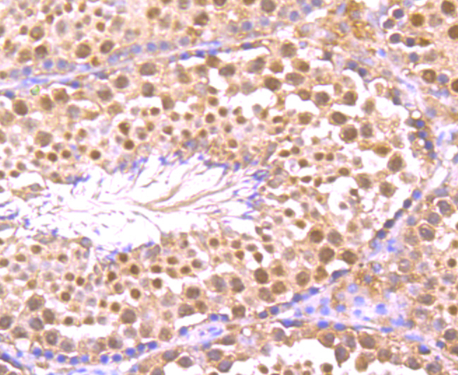
Fig7: Immunohistochemical analysis of paraffin-embedded mouse testis tissue using anti-TNFAIP3 antibody. Counter stained with hematoxylin.Application
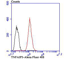
Fig8: Flow cytometric analysis of HepG2 cells with TNFAIP3 antibody at 1/100 dilution (red) compared with an unlabelled control (cells without incubation with primary antibody; black). Goat anti rabbit IgG (FITC) was used as the secondary antibody.| Product Name | TNFAIP3 |
|---|---|
| Antibody Type | Primary Antibodies |
| Antigen Alias | A20 antibody AISBL antibody MGC104522 antibody MGC138687 antibody MGC138688 antibody OTU domain containing protein 7C antibody OTU domain-containing protein 7C antibody OTUD7C antibody Putative DNA binding protein A20 antibody Putative DNA-binding protein A20 antibody TNAP3_HUMAN antibody TNF alpha-induced protein 3 antibody TNFA1P2 antibody TNFAIP 3 antibody TNFAIP3 (A20) antibody TNFAIP3 antibody Tumor necrosis factor alpha induced protein 3 antibody Tumor necrosis factor alpha-induced protein 3 antibody Tumor necrosis factor induced protein 3 antibody Tumor necrosis factor inducible protein A20 antibody tumor necrosis factor, alpha-induced protein 3 antibody Zinc finger protein A20 antibody |
| Product description | pecifically, A20 is induced by tumor necrosis factor (TNF) and interleukin 1 (IL-1), and acts as a negative regulator of nuclear factor κ B (NFκB) gene expression. By inhibiting NFκB activation, A20 plays a critical role in terminating NFκB responses to various stimuli. Although the C-terminal region of A20 contains seven zinc finger domains, only four of these domains are required for in vitro inhibition of TNF-induced NFκB activation. A20 also interacts with several other proteins, such as TRAF2, TRAF6 and IkB kinase (IKK) γ protein, and can thereby inhibit cell death. TXBP151, a novel A20-binding protein, may mediate the anti-apoptotic activity of A20. Involved in the negative feedback regulation of signal transduction, A20 and A20-binding proteins may be useful as novel therapeutic tools in the treatment of a variety of diseases. |
| Immunogen | Recombinant protein |
| Clonality | Polyclonal |
|---|---|
| Isotype | IgG |
| Host Species | Rabbit |
| Tested Applications | WBICCIHCFC |
| WB:1:500 ICC:1:100-1:500 IHC:1:50-1:200 FC:1:50-1:100 |
|
| Species Reactivity | HumanMouseRat |
| Concentration | 1mg/ml |
| Alternative Names | A20 antibody
AISBL antibody
MGC104522 antibody
MGC138687 antibody
MGC138688 antibody
OTU domain containing protein 7C antibody
OTU domain-containing protein 7C antibody
OTUD7C antibody
Putative DNA binding protein A20 antibody
Putative DNA-binding protein A20 antibody
TNAP3_HUMAN antibody
TNF alpha-induced protein 3 antibody
TNFA1P2 antibody
TNFAIP 3 antibody
TNFAIP3 (A20) antibody
TNFAIP3 antibody
Tumor necrosis factor alpha induced protein 3 antibody
Tumor necrosis factor alpha-induced protein 3 antibody
Tumor necrosis factor induced protein 3 antibody
Tumor necrosis factor inducible protein A20 antibody
tumor necrosis factor alpha-induced protein 3 antibody Zinc finger protein A20 antibody |
|---|---|
| Molecular Weight(MW) | 90 kDa |
| Cellular Localization | Cytoplasm. Nucleus. |
Application
Fig1: Western blot analysis of TNFAIP3 on Jurkat (1) and Daudi (2) cell lysate using anti-TNFAIP3 antibody at 1/100 dilution.
Application
Fig2: ICC staining TNFAIP3 in A431 cells (green). The nuclear counter stain is DAPI (blue). Cells were fixed in paraformaldehyde, permeabilised with 0.25% Triton X100/PBS.
Application
Fig3: ICC staining TNFAIP3 in LOVO cells (green). The nuclear counter stain is DAPI (blue). Cells were fixed in paraformaldehyde, permeabilised with 0.25% Triton X100/PBS.
Application
Fig4: Immunohistochemical analysis of paraffin-embedded rat lung tissue using anti-TNFAIP3 antibody. Counter stained with hematoxylin.
Application
Fig5: Immunohistochemical analysis of paraffin-embedded human kidney tissue using anti-TNFAIP3 antibody. Counter stained with hematoxylin.
Application
Fig6: Immunohistochemical analysis of paraffin-embedded human kidney tissue using anti-TNFAIP3 antibody. Counter stained with hematoxylin.
Application
Fig7: Immunohistochemical analysis of paraffin-embedded mouse testis tissue using anti-TNFAIP3 antibody. Counter stained with hematoxylin.
Application
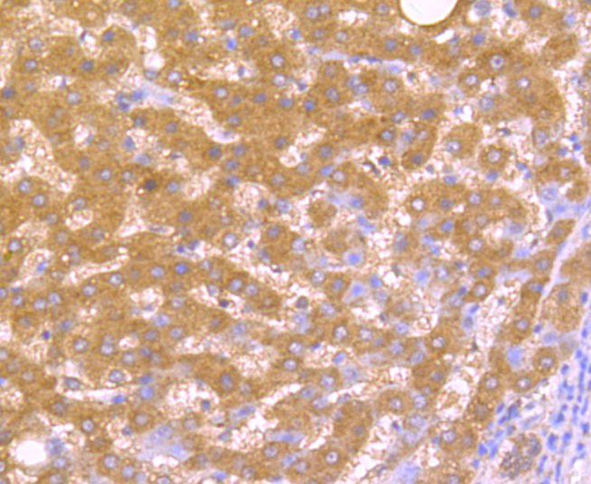
Fig8: Flow cytometric analysis of HepG2 cells with TNFAIP3 antibody at 1/100 dilution (red) compared with an unlabelled control (cells without incubation with primary antibody; black). Goat anti rabbit IgG (FITC) was used as the secondary antibody.| Positive Control | Jurkat, Daudi, A431, LOVO, rat lung tissue, human liver cancer tissue, human kidney tissue, mouse testis tissue, HepG2. |
|---|---|
| Application Notes | WB:1:500 ICC:1:100-1:500 IHC:1:50-1:200 FC:1:50-1:100 |
| Form | Liquid |
|---|---|
| Storage Instructions | Store at +4℃ after thawing. Aliquot store at -20℃ or -80℃. Avoid repeated freeze / thaw cycles. |
| Storage Buffer | 1*TBS (pH7.4), 0.5%BSA, 50%Glycerol. Preservative: 0.05% Sodium Azide. |


