Application
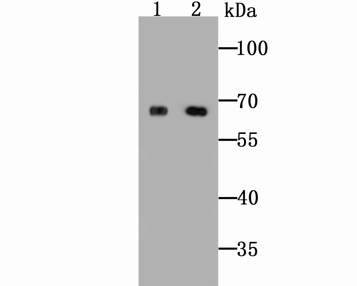
Fig1: Western blot analysis of ZIP12 on HepG2 cell and rat lung tissue lysates using anti-ZIP12 antibody at 1/500 dilution.Application
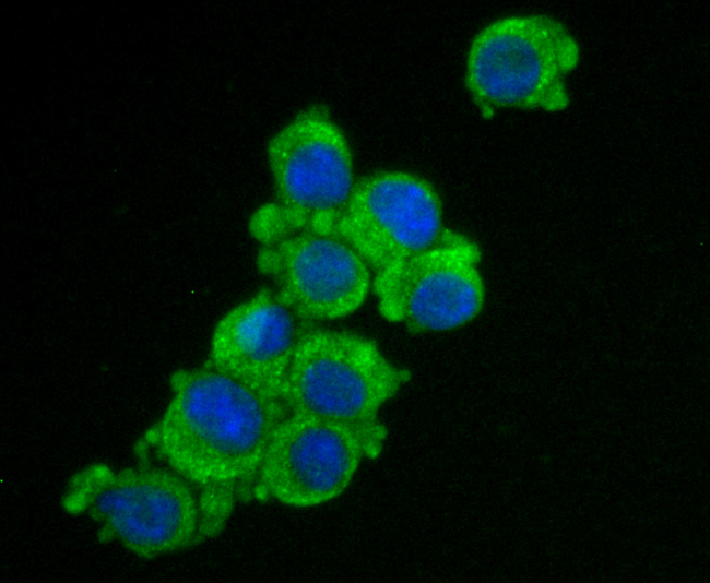
Fig2: ICC staining ZIP12 in N2A cells (green). The nuclear counter stain is DAPI (blue). Cells were fixed in paraformaldehyde, permeabilised with 0.25% Triton X100/PBS.Application
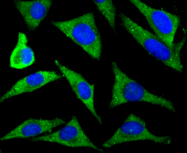
Fig3: ICC staining ZIP12 in SH-SY5Y cells (green). The nuclear counter stain is DAPI (blue). Cells were fixed in paraformaldehyde, permeabilised with 0.25% Triton X100/PBS.Application
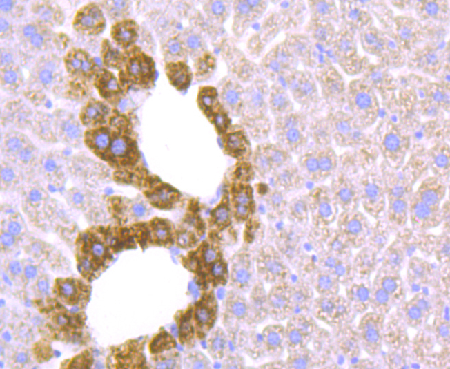
Fig4: Immunohistochemical analysis of paraffin-embedded mouse liver tissue using anti-ZIP12 antibody. Counter stained with hematoxylin.Application
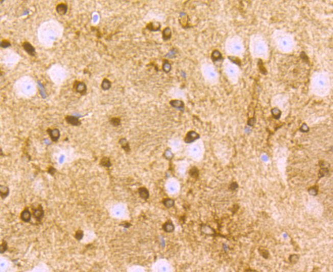
Fig5: Immunohistochemical analysis of paraffin-embedded mouse brain tissue using anti-ZIP12 antibody. Counter stained with hematoxylin.Application
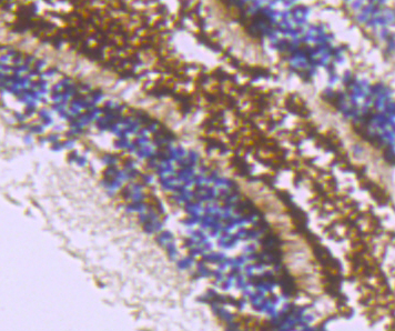
Fig6: Immunohistochemical analysis of paraffin-embedded mouse eyeball tissue using anti-ZIP12 antibody. Counter stained with hematoxylin.| Product Name | ZIP12 |
|---|---|
| Antibody Type | Primary Antibodies |
| Antigen Alias | bA570F3.1 antibody FLJ30499 antibody LIV 1 subfamily of ZIP zinc transporter 8 antibody LIV-1 subfamily of ZIP zinc transporter 8 antibody LZT Hs8 antibody LZT-Hs8 antibody LZTHs8 antibody MGC43205 antibody MGC51099 antibody S39AC_HUMAN antibody SLC39A12 antibody Solute carrier family 39 (metal ion transporter) member 12 antibody Solute carrier family 39 (zinc transporter) member 12 antibody Solute carrier family 39 member 12 antibody Zinc transporter ZIP12 antibody ZIP 12 antibody ZIP-12 antibody ZIP12 antibody Zrt and Irt like protein 12 antibody Zrt- and Irt-like protein 12 antibody |
| Product description | Acts as a zinc-influx transporter (Potential). May be partly involved in the outbreak of schizophrenia. On the basis of sequence homology, the human SLC39A12 gene has been included as a member of a family of zinc (Zn) transporters designated as the ZRT-IRT-like proteins (current designation SLC39). ZRT-IRT-like proteins act in concert with another family of transporters, known in non-mammalian organisms as cation diffusion facilitators and in mammals as zinc transporters (ZnT), to regulate the distribution of Zn in the extra- versus intracellular milieu and within cellular compartments. Hence the ZnT maintain Zn homeostasis that is critical for the normal functioning of tissues including the CNS. |
| Immunogen | Peptide |
| Clonality | Polyclonal |
|---|---|
| Isotype | IgG |
| Host Species | Rabbit |
| Tested Applications | WBICCIHC |
| WB:1:500-1:1,000 ICC:1:50-1:200 IHC:1:50-1:200 |
|
| Species Reactivity | HumanMouseRat |
| Concentration | 1mg/ml |
| Alternative Names | bA570F3.1 antibody FLJ30499 antibody LIV 1 subfamily of ZIP zinc transporter 8 antibody LIV-1 subfamily of ZIP zinc transporter 8 antibody LZT Hs8 antibody LZT-Hs8 antibody LZTHs8 antibody MGC43205 antibody MGC51099 antibody S39AC_HUMAN antibody SLC39A12 antibody Solute carrier family 39 (metal ion transporter) member 12 antibody Solute carrier family 39 (zinc transporter) member 12 antibody Solute carrier family 39 member 12 antibody Zinc transporter ZIP12 antibody ZIP 12 antibody ZIP-12 antibody ZIP12 antibody Zrt and Irt like protein 12 antibody Zrt- and Irt-like protein 12 antibody |
|---|---|
| Molecular Weight(MW) | 70 kDa |
| Cellular Localization | Membrane. |
Application
Fig1: Western blot analysis of ZIP12 on HepG2 cell and rat lung tissue lysates using anti-ZIP12 antibody at 1/500 dilution.
Application
Fig2: ICC staining ZIP12 in N2A cells (green). The nuclear counter stain is DAPI (blue). Cells were fixed in paraformaldehyde, permeabilised with 0.25% Triton X100/PBS.
Application
Fig3: ICC staining ZIP12 in SH-SY5Y cells (green). The nuclear counter stain is DAPI (blue). Cells were fixed in paraformaldehyde, permeabilised with 0.25% Triton X100/PBS.
Application
Fig4: Immunohistochemical analysis of paraffin-embedded mouse liver tissue using anti-ZIP12 antibody. Counter stained with hematoxylin.
Application
Fig5: Immunohistochemical analysis of paraffin-embedded mouse brain tissue using anti-ZIP12 antibody. Counter stained with hematoxylin.
Application
Fig6: Immunohistochemical analysis of paraffin-embedded mouse eyeball tissue using anti-ZIP12 antibody. Counter stained with hematoxylin.| Positive Control | HepG2, N2A, SH-SY5Y, rat lung tissue, mouse liver tissue, mouse brain tissue, mouse eyeball tissue. |
|---|---|
| Application Notes | WB:1:500-1:1,000 ICC:1:50-1:200 IHC:1:50-1:200 |
| Form | Liquid |
|---|---|
| Storage Instructions | Store at +4℃ after thawing. Aliquot store at -20℃ or -80℃. Avoid repeated freeze / thaw cycles. |
| Storage Buffer | 1*TBS (pH7.4), 0.5%BSA, 50%Glycerol. Preservative: 0.05% Sodium Azide. |


