Application
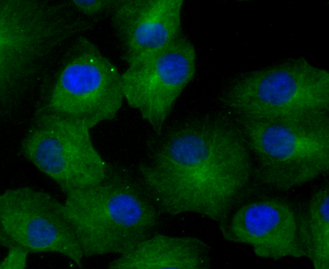
Fig1:ICC staining GCN2 in A549 cells (green). The nuclear counter stain is DAPI (blue). Cells were fixed in paraformaldehyde, permeabilised with 0.25% Triton X100/PBS.Application
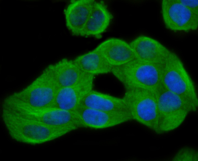
Fig2: ICC staining GCN2 in Hela cells (green). The nuclear counter stain is DAPI (blue). Cells were fixed in paraformaldehyde, permeabilised with 0.25% Triton X100/PBS.Application
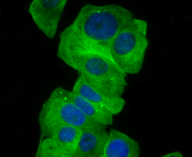
Fig3: ICC staining GCN2 in MCF-7 cells (green). The nuclear counter stain is DAPI (blue). Cells were fixed in paraformaldehyde, permeabilised with 0.25% Triton X100/PBS.Application
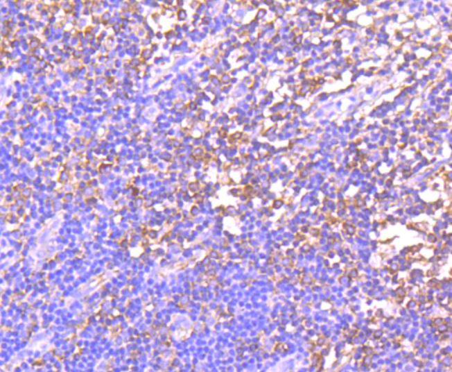
Fig4: Immunohistochemical analysis of paraffin-embedded human tonsil tissue using anti-GCN2 antibody. Counter stained with hematoxylin.Application
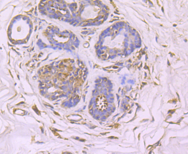
Fig5: Immunohistochemical analysis of paraffin-embedded human breast cancer tissue using anti-GCN2 antibody. Counter stained with hematoxylin.Application
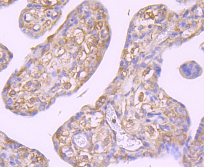
Fig6: Immunohistochemical analysis of paraffin-embedded human placenta tissue using anti-GCN2 antibody. Counter stained with hematoxylin.Application
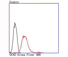
Fig7: Flow cytometric analysis of MCF-7 cells with GCN2 antibody at 1/100 dilution (red) compared with an unlabelled control (cells without incubation with primary antibody; black).| Product Name | GCN2 [JA03-83] |
|---|---|
| Antibody Type | Primary Antibodies |
| Antigen Alias | E2AK4_HUMAN antibody Eif2ak4 antibody Eukaryotic Translation Initiation Factor 2 alpha kinase 4 antibody Eukaryotic translation initiation factor 2-alpha kinase 4 antibody GCN2 antibody GCN2 eIF2alpha kinase antibody GCN2 like protein antibody GCN2-like protein antibody KIAA1338 antibody MGCN2 antibody |
| Product description | The family of stress-responsive protein kinases include HRI (heme-regulated inhibitor or EIF2AK1), PKR (EIF2AK2 or TIK), PERK (EIF2AK3) and GCN2 (EIF2AK4). These proteins phosphorylate the eukaryotic translation initiation factor 2α (eIF2α) on Ser 51 to regulate general and gene-specific protein synthesis. Phosphorylated eIF2α acts as an inhibitor of its guanine nucleotide exchange factor eIF2B. GCN2, a unique eIF2α kinase, exists in all eukaryotes from yeast to mammals. In mammals, expression of GCN2 is highest in liver and brain tissues. GCN2 primarily initiates the phosphorylation of eIF2α in response to UV, but has been shown to increase phosphorylation activity in response to serum starvation. Also, substitution of Asp 83 for Ala on eIF2α results in impaired phosphorylation by GCN2 and PKR, suggesting a contribution of remote residues to kinase-substrate recognition. |
| Immunogen | recombinant protein |
| Clonality | Monoclonal |
|---|---|
| Isotype | IgG |
| Host Species | Recombinant rabbit |
| Tested Applications | WBICCIHCFC |
| WB:1:500-1:1,000 ICC:1:50-1:200 IHC:1:50-1:200 FC:1:50-1:100 |
|
| Species Reactivity | Human |
| Concentration | 1mg/ml |
| Alternative Names | E2AK4_HUMAN antibody Eif2ak4 antibody Eukaryotic Translation Initiation Factor 2 alpha kinase 4 antibody Eukaryotic translation initiation factor 2-alpha kinase 4 antibody GCN2 antibody GCN2 eIF2alpha kinase antibody GCN2 like protein antibody GCN2-like protein antibody KIAA1338 antibody MGCN2 antibody |
|---|---|
| Molecular Weight(MW) | 187 kDa |
| Cellular Localization | Cytoplasm. |
| SwissProt ID | Q9P2K8 |
|---|
Application
Fig1:ICC staining GCN2 in A549 cells (green). The nuclear counter stain is DAPI (blue). Cells were fixed in paraformaldehyde, permeabilised with 0.25% Triton X100/PBS.
Application
Fig2: ICC staining GCN2 in Hela cells (green). The nuclear counter stain is DAPI (blue). Cells were fixed in paraformaldehyde, permeabilised with 0.25% Triton X100/PBS.
Application
Fig3: ICC staining GCN2 in MCF-7 cells (green). The nuclear counter stain is DAPI (blue). Cells were fixed in paraformaldehyde, permeabilised with 0.25% Triton X100/PBS.
Application
Fig4: Immunohistochemical analysis of paraffin-embedded human tonsil tissue using anti-GCN2 antibody. Counter stained with hematoxylin.
Application
Fig5: Immunohistochemical analysis of paraffin-embedded human breast cancer tissue using anti-GCN2 antibody. Counter stained with hematoxylin.
Application
Fig6: Immunohistochemical analysis of paraffin-embedded human placenta tissue using anti-GCN2 antibody. Counter stained with hematoxylin.
Application
Fig7: Flow cytometric analysis of MCF-7 cells with GCN2 antibody at 1/100 dilution (red) compared with an unlabelled control (cells without incubation with primary antibody; black).| Positive Control | A549, Hela, MCF-7, human tonsil tissue, human breast cancer tissue, human placenta tissue. |
|---|---|
| Application Notes | WB:1:500-1:1,000 ICC:1:50-1:200 IHC:1:50-1:200 FC:1:50-1:100 |
| Form | Liquid |
|---|---|
| Storage Instructions | Store at +4℃ after thawing. Aliquot store at -20℃ or -80℃. Avoid repeated freeze / thaw cycles. |
| Storage Buffer | 1*TBS (pH7.4), 1%BSA, 40%Glycerol. Preservative: 0.05% Sodium Azide. |


