Application
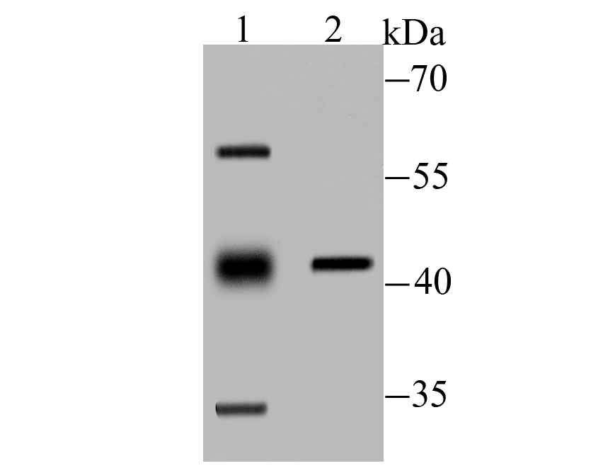
Fig1: Western blot analysis of PP2C alpha on mouse kidney tissue (1) and rat skin tissue (2) lysate using anti-PP2C alpha antibody at 1/100 dilution.Application
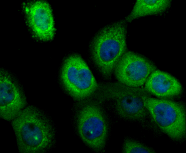
Fig2: ICC staining PP2C alpha in A431 cells (green). The nuclear counter stain is DAPI (blue). Cells were fixed in paraformaldehyde, permeabilised with 0.25% Triton X100/PBS.Application
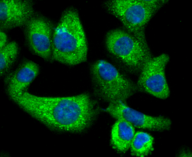
Fig3: ICC staining PP2C alpha in A549 cells (green). The nuclear counter stain is DAPI (blue). Cells were fixed in paraformaldehyde, permeabilised with 0.25% Triton X100/PBS.Application
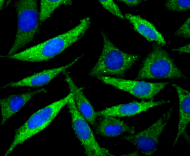
Fig4: ICC staining PP2C alpha in SH-SY5Y cells (green). The nuclear counter stain is DAPI (blue). Cells were fixed in paraformaldehyde, permeabilised with 0.25% Triton X100/PBS.Application
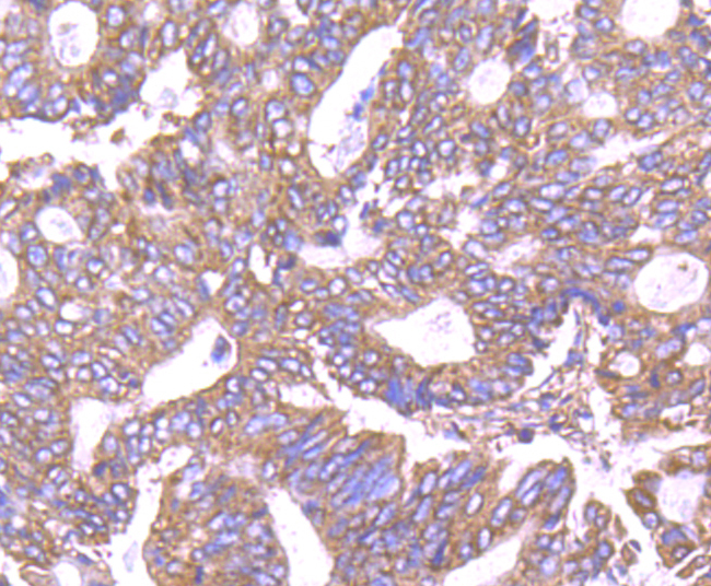
Fig5: Immunohistochemical analysis of paraffin-embedded human colon cancer tissue using anti-PP2C alpha antibody. Counter stained with hematoxylin.Application
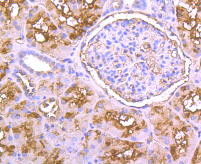
Fig6: Immunohistochemical analysis of paraffin-embedded human kidney tissue using anti-PP2C alpha antibody. Counter stained with hematoxylin.Application
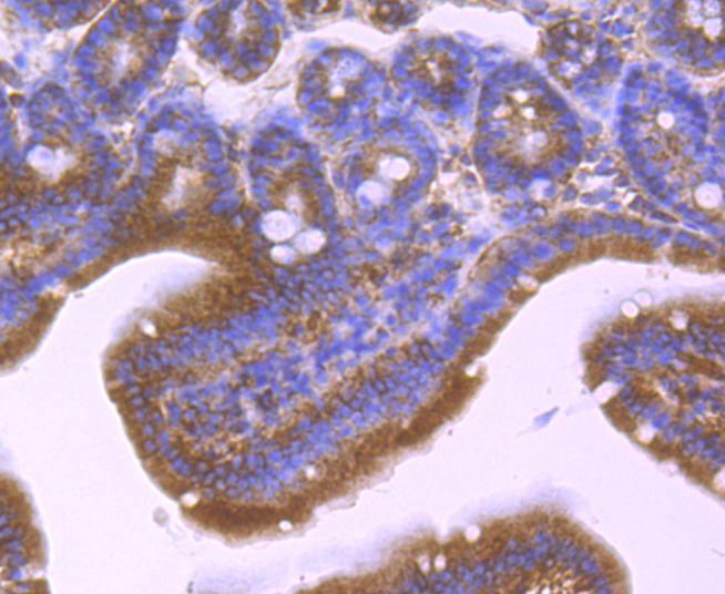
Fig7: Immunohistochemical analysis of paraffin-embedded mouse small intestine tissue using anti-PP2C alpha antibody. Counter stained with hematoxylin.Application
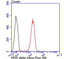
Fig8: Flow cytometric analysis of A431 cells with PP2C alpha antibody at 1/100 dilution (red) compared with an unlabelled control (cells without incubation with primary antibody; black). Alexa Fluor 488-conjugated goat anti rabbit IgG was used as the secondary antibody.| Product Name | PP2C alpha |
|---|---|
| Antibody Type | Primary Antibodies |
| Antigen Alias | Protein phosphatase 2C isoform alpha antibody EC 3.1.3.16 antibody FLJ42306 antibody IA antibody MGC9201 antibody Mpp alpha antibody PP2C alpha antibody PP2C-alpha antibody PP2CA antibody PP2Calpha antibody PPM 1A antibody PPM1A antibody PPM1A_HUMAN antibody PPPM1A antibody Protein phosphatase 1A (formerly 2C) magnesium dependent alpha isoform antibody Protein phosphatase 1A antibody Protein phosphatase 1A magnesium dependent alpha antibody Protein phosphatase 2C alpha antibody Protein phosphatase 2C alpha isoform antibody Protein phosphatase 2C isoform alpha antibody Protein phosphatase IA antibody Protein phosphatase, Mg2+/Mn2+ dependent, 1A antibody |
| Product description | The phosphorylation and dephosphorylation of proteins on serine and threonine residues is an essential means of regulating a broad range of cellular functions in eukaryotes, including division, homeostasis and apoptosis. A group of proteins that are intimately involved in this process are the serine/ threonine protein phosphatases. Protein phosphatase 2Ca (PP2Ca) has broad specificity. It dephosphorylates and negatively regulates the activities of MAP kinases and MAP kinase-kinases while also inhibiting the activation of p38 and JNK kinase cascades, induced by environmental stresses. PP2Ca also induces the expression of endogenous p53 and the p53-responsive gene p21, leading to cell cycle arrest and apoptosis. The PP2Ca protein, which contains an active site containing a dinuclear metal ion center, shows highest expression in epithelial cells, as well as in the digestive tract, lung, kidney, breast, prostate, endocrine glands and brain. |
| Immunogen | Recombinant protein |
| Clonality | Polyclonal |
|---|---|
| Isotype | IgG |
| Host Species | Rabbit |
| Tested Applications | WBICCIHCFC |
| WB:1:500 ICC:1:100-1:500 IHC:1:50-1:200 FC:1:50-1:100 |
|
| Species Reactivity | HumanMouseRat |
| Concentration | 1mg/ml |
| Alternative Names | Protein phosphatase 2C isoform alpha antibody
EC 3.1.3.16 antibody
FLJ42306 antibody IA antibody
MGC9201 antibody
Mpp alpha antibody
PP2C alpha antibody
PP2C-alpha antibody
PP2CA antibody
PP2Calpha antibody
PPM 1A antibody
PPM1A antibody
PPM1A_HUMAN antibody
PPPM1A antibody
Protein phosphatase 1A (formerly 2C) magnesium dependent alpha isoform antibody
Protein phosphatase 1A antibody
Protein phosphatase 1A magnesium dependent alpha antibody
Protein phosphatase 2C alpha antibody
Protein phosphatase 2C alpha isoform antibody
Protein phosphatase 2C isoform alpha antibody
Protein phosphatase IA antibody
Protein phosphatase Mg2+/Mn2+ dependent 1A antibody |
|---|---|
| Molecular Weight(MW) | 42/35 kDa, additional band 55 kDa. |
| Cellular Localization | Cytoplasm, Membrane, Nucleus. |
Application
Fig1: Western blot analysis of PP2C alpha on mouse kidney tissue (1) and rat skin tissue (2) lysate using anti-PP2C alpha antibody at 1/100 dilution.
Application
Fig2: ICC staining PP2C alpha in A431 cells (green). The nuclear counter stain is DAPI (blue). Cells were fixed in paraformaldehyde, permeabilised with 0.25% Triton X100/PBS.
Application
Fig3: ICC staining PP2C alpha in A549 cells (green). The nuclear counter stain is DAPI (blue). Cells were fixed in paraformaldehyde, permeabilised with 0.25% Triton X100/PBS.
Application
Fig4: ICC staining PP2C alpha in SH-SY5Y cells (green). The nuclear counter stain is DAPI (blue). Cells were fixed in paraformaldehyde, permeabilised with 0.25% Triton X100/PBS.
Application
Fig5: Immunohistochemical analysis of paraffin-embedded human colon cancer tissue using anti-PP2C alpha antibody. Counter stained with hematoxylin.
Application
Fig6: Immunohistochemical analysis of paraffin-embedded human kidney tissue using anti-PP2C alpha antibody. Counter stained with hematoxylin.
Application
Fig7: Immunohistochemical analysis of paraffin-embedded mouse small intestine tissue using anti-PP2C alpha antibody. Counter stained with hematoxylin.
Application
Fig8: Flow cytometric analysis of A431 cells with PP2C alpha antibody at 1/100 dilution (red) compared with an unlabelled control (cells without incubation with primary antibody; black). Alexa Fluor 488-conjugated goat anti rabbit IgG was used as the secondary antibody.| Positive Control | Mouse kidney tissue lysate, rat skin tissue lysate, A431, A549, SH-SY5Y, human kidney tissue, human colon cancer tissue, mouse intestine tissue. |
|---|---|
| Application Notes | WB:1:500 ICC:1:100-1:500 IHC:1:50-1:200 FC:1:50-1:100 |
| Form | Liquid |
|---|---|
| Storage Instructions | Store at +4℃ after thawing. Aliquot store at -20℃ or -80℃. Avoid repeated freeze / thaw cycles. |
| Storage Buffer | 1*TBS (pH7.4), 0.5%BSA, 50%Glycerol. Preservative: 0.05% Sodium Azide. |


