Application
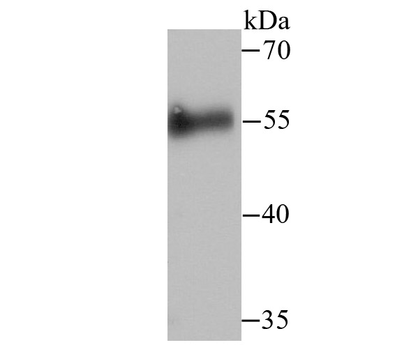
Fig1: Western blot analysis of Cdc25C on human skin tissue lysate using anti-Cdc25C antibody at 1/500 dilution.Application
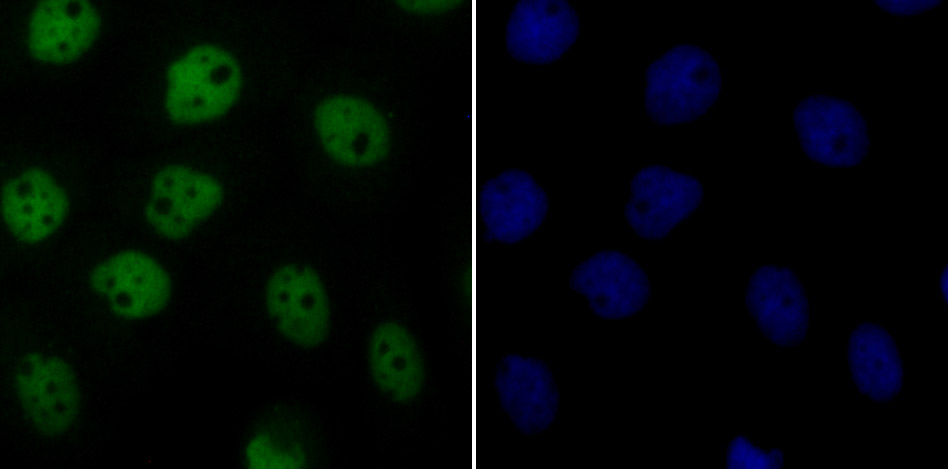
Fig2: ICC staining Cdc25C in A431 cells (green). The nuclear counter stain is DAPI (blue). Cells were fixed in paraformaldehyde, permeabilised with 0.25% Triton X100/PBS.Application
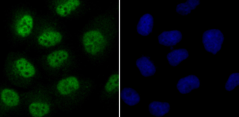
Fig3: ICC staining Cdc25C in HUVEC cells (green). The nuclear counter stain is DAPI (blue). Cells were fixed in paraformaldehyde, permeabilised with 0.25% Triton X100/PBS.Application
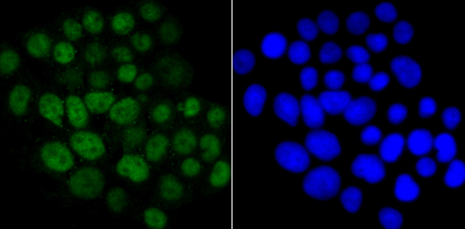
Fig4: ICC staining Cdc25C in LOVO cells (green). The nuclear counter stain is DAPI (blue). Cells were fixed in paraformaldehyde, permeabilised with 0.25% Triton X100/PBS.Application
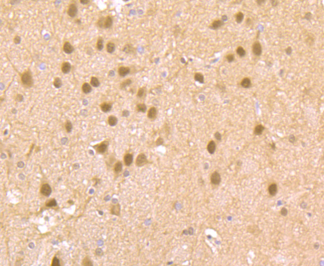
Fig5: Immunohistochemical analysis of paraffin-embedded rat brain tissue using anti-Cdc25C antibody. Counter stained with hematoxylin.Application
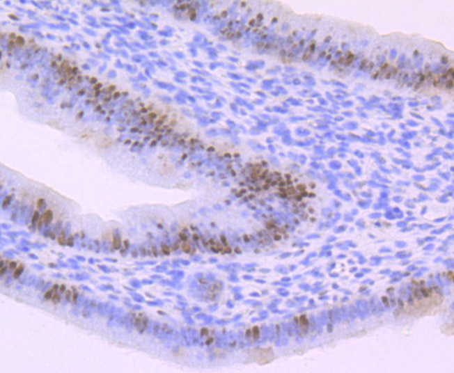
Fig6: Immunohistochemical analysis of paraffin-embedded mouse uterus tissue using anti-Cdc25C antibody. Counter stained with hematoxylin.Application
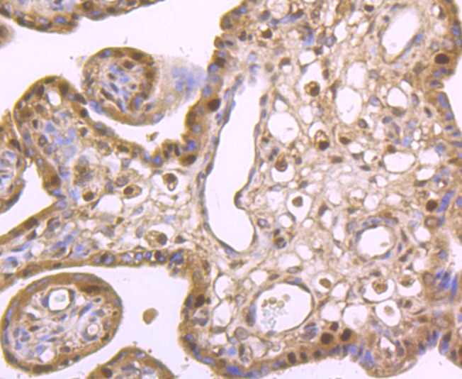
Fig7: Immunohistochemical analysis of paraffin-embedded human placenta tissue using anti-Cdc25C antibody. Counter stained with hematoxylin.Application
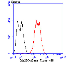
Fig8: Flow cytometric analysis of LOVO cells with Cdc25C antibody at 1/100 dilution (red) compared with an unlabelled control (cells without incubation with primary antibody; black). Alexa Fluor 488-conjugated goat anti rabbit IgG was used as the secondary antibody.| Product Name | Cdc25C |
|---|---|
| Antibody Type | Primary Antibodies |
| Antigen Alias | CDC 25 antibody Cdc 25C antibody CDC25 antibody CDC25C antibody Cell division cycle 25 homolog C antibody Cell division cycle 25C antibody Cell division cycle 25C protein antibody Dual specificity phosphatase Cdc25C antibody M phase inducer phosphatase 3 antibody M-phase inducer phosphatase 3 antibody Mitosis inducer CDC25 antibody MPIP3 antibody MPIP3_HUMAN antibody Phosphotyrosine phosphatase antibody PPP1R60 antibody protein phosphatase 1, regulatory subunit 60 antibody |
| Product description | Cell cycle events are regulated by the sequential activation and deactivation of cyclin-dependent kinases (Cdks), including Cdk2 and Cdc2. Cdk2 in complexes with cyclin E and cyclin A appears necessary for the onset and progression of DNA replication, while the Cdc2 kinase in complexes with cyclin A or cyclin B is required for the initiation of cell division. Wee 1 has been identified as a protein kinase that suppresses the entry into mitosis by mediating inhibiting tyrosine phosphorylation of Cdc2 p34. In contrast, members of the Cdc25 family of protein phosphatases function as mitotic activators by dephosphorylation of Cdc2 p34 on regulatory tyrosine and possibly threonine residues. The Cdc25 gene family consists of at least three members that share approximately 40% identity in their most conserved carboxy-terminal sequences. |
| Immunogen | Peptide |
| Clonality | Polyclonal |
|---|---|
| Isotype | IgG |
| Host Species | Rabbit |
| Tested Applications | WBICCIHCFC |
| WB:1:500-1:1,000 ICC:1:500-1:2,000 IHC:1:50-1:200 FC:1:50-1:100 |
|
| Species Reactivity | HumanMouseRat |
| Concentration | 1mg/ml |
| Alternative Names | CDC 25 antibody
Cdc 25C antibody
CDC25 antibody
CDC25C antibody
Cell division cycle 25 homolog C antibody
Cell division cycle 25C antibody
Cell division cycle 25C protein antibody
Dual specificity phosphatase Cdc25C antibody
M phase inducer phosphatase 3 antibody
M-phase inducer phosphatase 3 antibody
Mitosis inducer CDC25 antibody
MPIP3 antibody
MPIP3_HUMAN antibody
Phosphotyrosine phosphatase antibody
PPP1R60 antibody
protein phosphatase 1 regulatory subunit 60 antibody |
|---|---|
| Molecular Weight(MW) | 53 kDa |
| Cellular Localization | Nucleus. |
Application
Fig1: Western blot analysis of Cdc25C on human skin tissue lysate using anti-Cdc25C antibody at 1/500 dilution.
Application
Fig2: ICC staining Cdc25C in A431 cells (green). The nuclear counter stain is DAPI (blue). Cells were fixed in paraformaldehyde, permeabilised with 0.25% Triton X100/PBS.
Application
Fig3: ICC staining Cdc25C in HUVEC cells (green). The nuclear counter stain is DAPI (blue). Cells were fixed in paraformaldehyde, permeabilised with 0.25% Triton X100/PBS.
Application
Fig4: ICC staining Cdc25C in LOVO cells (green). The nuclear counter stain is DAPI (blue). Cells were fixed in paraformaldehyde, permeabilised with 0.25% Triton X100/PBS.
Application
Fig5: Immunohistochemical analysis of paraffin-embedded rat brain tissue using anti-Cdc25C antibody. Counter stained with hematoxylin.
Application
Fig6: Immunohistochemical analysis of paraffin-embedded mouse uterus tissue using anti-Cdc25C antibody. Counter stained with hematoxylin.
Application
Fig7: Immunohistochemical analysis of paraffin-embedded human placenta tissue using anti-Cdc25C antibody. Counter stained with hematoxylin.
Application
Fig8: Flow cytometric analysis of LOVO cells with Cdc25C antibody at 1/100 dilution (red) compared with an unlabelled control (cells without incubation with primary antibody; black). Alexa Fluor 488-conjugated goat anti rabbit IgG was used as the secondary antibody.| Positive Control | Human skin tissue lysate, A431, HUVEC, LOVO, rat brain tissue, human placenta tissue, mouse uterus tissue. |
|---|---|
| Application Notes | WB:1:500-1:1,000 ICC:1:500-1:2,000 IHC:1:50-1:200 FC:1:50-1:100 |
| Form | Liquid |
|---|---|
| Storage Instructions | Store at +4℃ after thawing. Aliquot store at -20℃ or -80℃. Avoid repeated freeze / thaw cycles. |
| Storage Buffer | 1*TBS (pH7.4), 0.5%BSA, 50%Glycerol. Preservative: 0.05% Sodium Azide. |


