Application
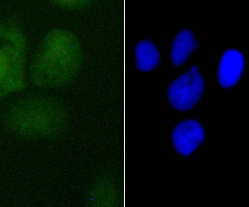
Fig1: ICC staining PDCD4 in A431 cells (green). The nuclear counter stain is DAPI (blue). Cells were fixed in paraformaldehyde, permeabilised with 0.25% Triton X100/PBS.Application
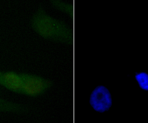
Fig2: ICC staining PDCD4 in PC-3M cells (green). The nuclear counter stain is DAPI (blue). Cells were fixed in paraformaldehyde, permeabilised with 0.25% Triton X100/PBS.Application
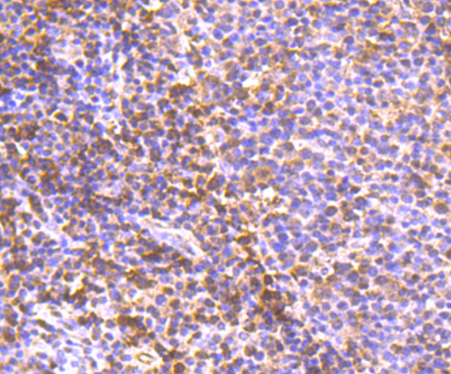
Fig3: Immunohistochemical analysis of paraffin-embedded human tonsil tissue using anti-PDCD4 antibody. Counter stained with hematoxylin.Application
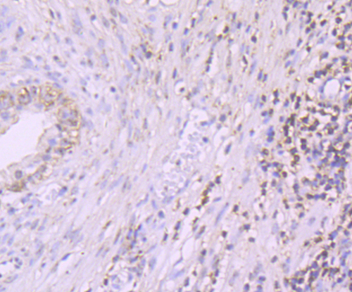
Fig4: Immunohistochemical analysis of paraffin-embedded human prostate tissue using anti-PDCD4 antibody. Counter stained with hematoxylin.Application
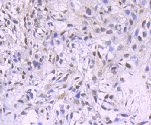
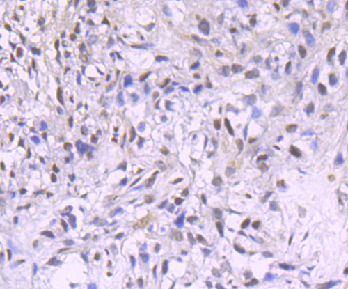
Fig5: Immunohistochemical analysis of paraffin-embedded human breast cancer tissue using anti-PDCD4 antibody. Counter stained with hematoxylin.Application
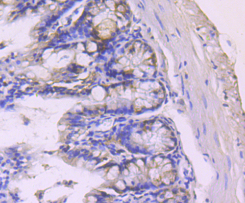
Fig6: Immunohistochemical analysis of paraffin-embedded mouse colon tissue using anti-PDCD4 antibody. Counter stained with hematoxylin.Application
Fig7: Flow cytometric analysis of MCF-7 cells with PDCD4 antibody at 1/100 dilution (red) compared with an unlabelled control (cells without incubation with primary antibody; black).| Product Name | PDCD4 [JA80-83] |
|---|---|
| Antibody Type | Primary Antibodies |
| Antigen Alias | Death up-regulated gene protein antibody Dug antibody H731 antibody Ma3 antibody MGC33046 antibody MGC33047 antibody Neoplastic transformation inhibitor antibody Neoplastic transformation inhibitor protein antibody Nuclear antigen H731 antibody Nuclear antigen H731 like antibody Nuclear antigen H731 like protein antibody Nuclear antigen H731-like antibody PDCD 4 antibody Pdcd4 antibody PDCD4_HUMAN antibody Programmed cell death 4 antibody programmed cell death 4 (neoplastic transformation inhibitor) antibody Programmed cell death protein 4 antibody Protein 197/15a antibody Protein MA-3 antibody Tis antibody Topoisomerase-inhibitor suppressed protein antibody |
| Product description | The transformation suppressor gene Pdcd-4 (programmed cell death gene 4) inhibits the tumor-promoter mediated transformation of mouse keratinocytes and is a potential tumor suppressor gene in the development of human lung cancer. Biochemical analysis suggests that the Pdcd-4 protein is involved in protein translation as well as in nuclear events. Pdcd-4 directly interacts with the RNA helicase eIF4A and inhibits protein synthesis by interfering with the assembly of the cap-dependent translation initiation complex. Pdcd-4 also suppresses the transactivation of AP-1 responsive promoters by c-Jun, suggesting that the transformation-suppressor activity of Pdcd-4 might be due, at least in part, to the inhibition of c-Jun activity. In addition to affecting c-Jun phosphorylation, Pdcd-4 blocks the recruitment of the co-activator p300 by c-Jun. |
| Immunogen | Recombinant protein |
| Clonality | Monoclonal |
|---|---|
| Isotype | IgG |
| Host Species | Recombinant rabbit |
| Tested Applications | WBICCIHCFC |
| WB:1:500~1:2000 ICC:1:200~1:500 IHC:1:50~1:200 FC:1:200~1:500 Notes:Optimal dilutions/concentrations should be determined by the researcher. |
|
| Species Reactivity | HumanMouse |
| Concentration | 1mg/ml |
| Alternative Names | Death up-regulated gene protein antibody Dug antibody H731 antibody Ma3 antibody MGC33046 antibody MGC33047 antibody Neoplastic transformation inhibitor antibody Neoplastic transformation inhibitor protein antibody Nuclear antigen H731 antibody Nuclear antigen H731 like antibody Nuclear antigen H731 like protein antibody Nuclear antigen H731-like antibody PDCD 4 antibody Pdcd4 antibody PDCD4_HUMAN antibody Programmed cell death 4 antibody programmed cell death 4 (neoplastic transformation inhibitor) antibody Programmed cell death protein 4 antibody Protein 197/15a antibody Protein MA-3 antibody Tis antibody Topoisomerase-inhibitor suppressed protein antibody |
|---|---|
| Molecular Weight(MW) | 52 kDa |
| Cellular Localization | Nucleus. Cytoplasm. |
Application
Fig1: ICC staining PDCD4 in A431 cells (green). The nuclear counter stain is DAPI (blue). Cells were fixed in paraformaldehyde, permeabilised with 0.25% Triton X100/PBS.
Application
Fig2: ICC staining PDCD4 in PC-3M cells (green). The nuclear counter stain is DAPI (blue). Cells were fixed in paraformaldehyde, permeabilised with 0.25% Triton X100/PBS.
Application
Fig3: Immunohistochemical analysis of paraffin-embedded human tonsil tissue using anti-PDCD4 antibody. Counter stained with hematoxylin.
Application
Fig4: Immunohistochemical analysis of paraffin-embedded human prostate tissue using anti-PDCD4 antibody. Counter stained with hematoxylin.
Application
Fig5: Immunohistochemical analysis of paraffin-embedded human breast cancer tissue using anti-PDCD4 antibody. Counter stained with hematoxylin.
Application
Fig6: Immunohistochemical analysis of paraffin-embedded mouse colon tissue using anti-PDCD4 antibody. Counter stained with hematoxylin.
Application
Fig7: Flow cytometric analysis of MCF-7 cells with PDCD4 antibody at 1/100 dilution (red) compared with an unlabelled control (cells without incubation with primary antibody; black).| Positive Control | A431, PC-3M, Hmuan tonsil tissue, human prostate tissue, human breasr cancer tissue, mouse colon tissue, MCF-7. |
|---|---|
| Application Notes | WB:1:500~1:2000 ICC:1:200~1:500 IHC:1:50~1:200 FC:1:200~1:500 Notes:Optimal dilutions/concentrations should be determined by the researcher. |
| Form | Liquid |
|---|---|
| Storage Instructions | Store at +4℃ after thawing. Aliquot store at -20℃ or -80℃. Avoid repeated freeze / thaw cycles. |
| Storage Buffer | 1*TBS (pH7.4), 1%BSA, 40%Glycerol. Preservative: 0.05% Sodium Azide. |


